Review
. 2022 Jul;50(7):1027-1041.
doi: 10.1124/dmd.121.000478. Epub 2022 Feb 22.

From Steroid and Drug Metabolism to Glycobiology, Using Sulfotransferase Structures to Understand and Tailor Function

Affiliations
Review

From Steroid and Drug Metabolism to Glycobiology, Using Sulfotransferase Structures to Understand and Tailor Function

Lars C Pedersen et al. Drug Metab Dispos. 2022 Jul.

Abstract

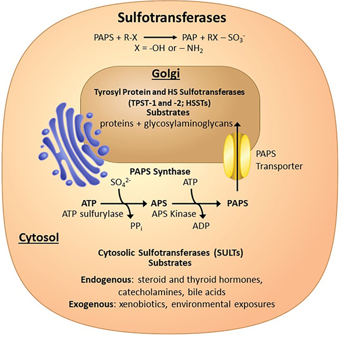
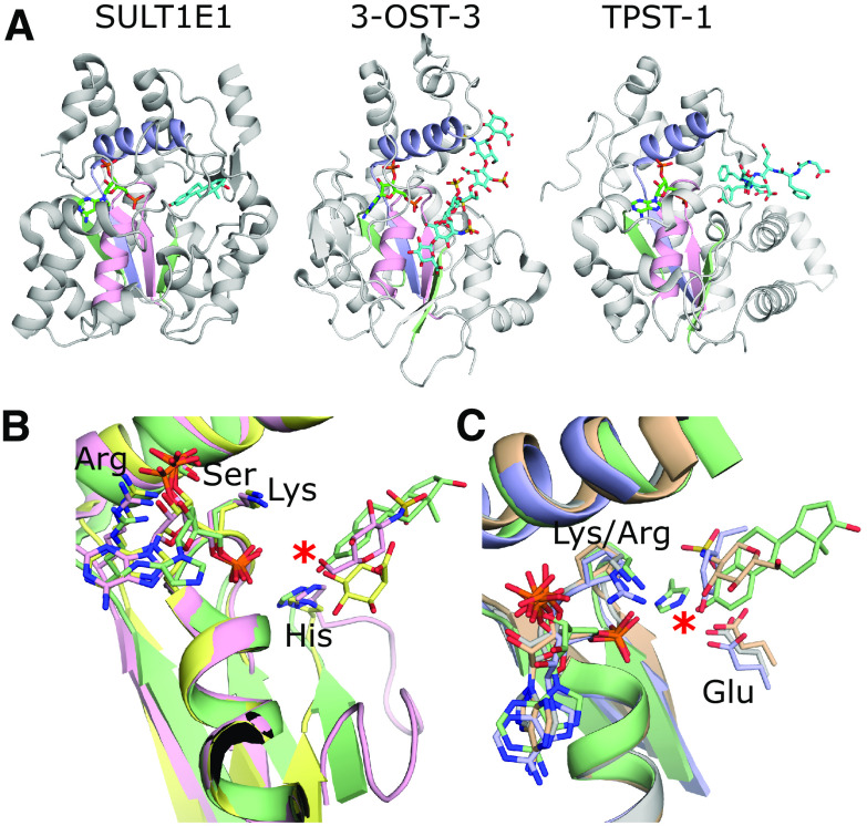
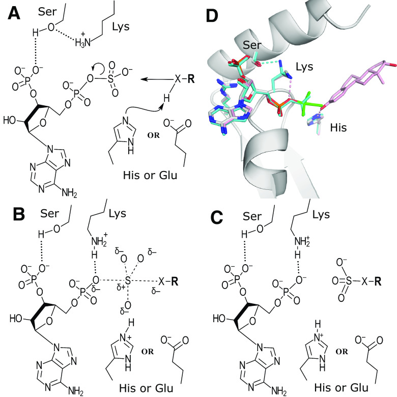
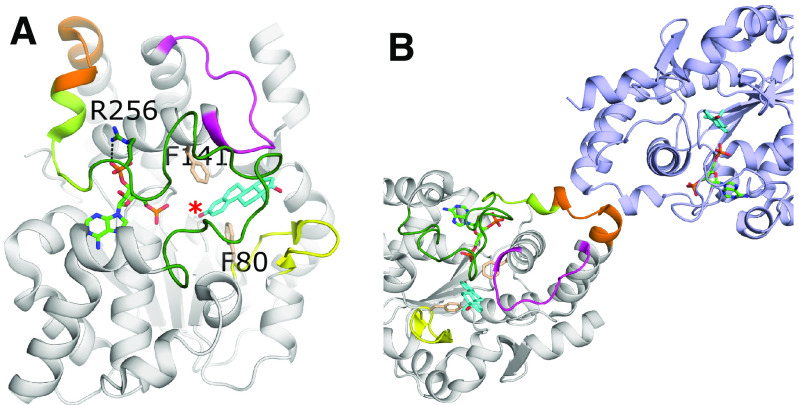
Sulfotransferases are ubiquitous enzymes that transfer a sulfo group from the universal cofactor donor 3'-phosphoadenosine 5'-phosphosulfate to a broad range of acceptor substrates. In humans, the cytosolic sulfotransferases are involved in the sulfation of endogenous compounds such as steroids, neurotransmitters, hormones, and bile acids as well as xenobiotics including drugs, toxins, and environmental chemicals. The Golgi associated membrane-bound sulfotransferases are involved in post-translational modification of macromolecules from glycosaminoglycans to proteins. The sulfation of small molecules can have profound biologic effects on the functionality of the acceptor, including activation, deactivation, or enhanced metabolism and elimination. Sulfation of macromolecules has been shown to regulate a number of physiologic and pathophysiological pathways by enhancing binding affinity to regulatory proteins or binding partners. Over the last 25 years, crystal structures of these enzymes have provided a wealth of information on the mechanisms of this process and the specificity of these enzymes. This review will focus on the general commonalities of the sulfotransferases, from enzyme structure to catalytic mechanism as well as providing examples into how structural information is being used to either design drugs that inhibit sulfotransferases or to modify the enzymes to improve drug synthesis. SIGNIFICANCE STATEMENT: This manuscript honors Dr. Masahiko Negishi's contribution to the understanding of sulfotransferase mechanism, specificity, and roles in biology by analyzing the crystal structures that have been solved over the last 25 years.


Figures

Fig. 1.
Fig. 1.
Sulfotransferases covered in this review. Sulfotransferases transfer a sulfo group (SO3) from PAPS, which is generated in the cytosol by the bifunctional PAPS synthases to many different types of acceptor substrates. The SULT enzymes in the cytosol sulfate endogenous and exogenous small molecules, whereas the Golgi-associated sulfotransferase sulfate macromolecules such as proteins and glycosaminoglycans.
Fig. 2.
Fig. 2.
Representative structures of the different types of sulfotransferases and their PAPS binding sites. (A) From left to right: crystal structure of hSULT1E1 in complex with PAP and estradiol [protein data bank (PDB) code 4JVL) (Gosavi et al., 2013); crystal structure of 3-OST-3 with PAP and an 8mer NS2S heparan sulfate bound (PDB code 6XL8) (Wander et al., 2021); crystal structure of TPST-1 with PAP and a polypeptide substrate bound (PDB code 5WRI) (Tanaka et al., 2017). The PAP is colored green and acceptor substrates are cyan. The strand-loop-helix containing the PSB loop is colored pink, whereas the strand-loop-helix containing the 3′-phosphate binding motif (3′SB) is colored light purple. The remaining two strands making up the central β-sheet are colored light green. (B) Comparisons of the PAP binding motifs and catalytic residues. The SULTs including SULT1E1 (green) and heparan sulfotransferases 2-OST (yellow) and 6-OST (pink) all use a lysine on the PSB loop and histidines for the proposed catalytic base (PDB codes 4JVL, 4NDZ, and 5T0A, respectively) (Gosavi et al., 2013; Liu et al., 2014; Xu et al., 2017b). The acceptor hydroxyl on the substrates all superimpose well, supporting a conserved mechanism. (C) Comparisons of the PAP binding motifs and catalytic residues of the sulfotransferase domain of SULT1E1 (green), NDST-1 (gray), 3-OST-3 (wheat), and TPST-1 (light purple) (PDB codes: 4JVL, NST1, 6XL8, and 5WRI, respectively) (Kakuta et al., 1999; Gosavi et al., 2013; Tanaka et al., 2017; Wander et al., 2021). NDST-1, 3-OST-3, and TPST-1 appear to use a conserved glutamate for their catalytic base, as opposed to the SULTs, 2-OST, and 6-OST. However, the PAPs and acceptor hydroxyls on the substrates all superimpose perfectly, supporting a conserved inline reaction mechanism for all the vertebrate sulfotransferases. For panels B and C, only the acceptor saccharide of the substrate is shown for 3-OST-3 and 2-OST and acceptor tyrosine of the peptide for TPST-1. No substrate is present in the crystal structure of NST-1.
Fig. 3.
Fig. 3.
Catalytic mechanism of the sulfotransferases. (A–C) Proposed catalytic mechanism of the PAPS-dependent sulfotransferases. (D) Common architecture of the sulfotransferase PAPS binding site including the PSB-loop and 3′SB strand-loop-helix from the crystal structure of mSULT1E1 (PDB code 1BO6) with PAP (white) and a vanadate molecule (green) in a trigonal bipyramidal arrangement thought to mimic the transition state. Also shown are the proposed catalytic base (His) and acid (Lys) (Kakuta et al., 1998b). Superimposed onto this structure are PAPS (cyan) and estradiol (pink) from two different structures of human SULT1E1 (PDB codes 1HY3 and 4JVL, respectively) with their equivalent His and Lys residues (Pedersen et al., 2002; Gosavi et al., 2013). When PAPS is present, the lysine forms a hydrogen bond with a conserved serine from the 3′SB binding region but is bound to the 5′-phosphate when PAP is present. These structures, combined with comparisons to uridylate kinase, shaped the hypothesis for the reaction mechanism shown in panels (A–C).
Fig. 4.
Fig. 4.
Substrate binding and dimerization of SULTS. (A) A representative SULT crystal structure, SULT1E1 with PAP (green) and estradiol (cyan) bound (PDB code 4JVL) (Gosavi et al., 2013). Shown are Loops 1 (yellow), 2 (magenta), and 3 (green) that contribute to substrate specificity and binding. The SULT-specific GXXGXXK region that connects loop 3 to the dimerization domain (orange) is colored lemon. The acceptor hydroxyl of the estradiol is designated with a red asterisk. Also shown are highly conserved aromatic residues (wheat) found in the SULT1 enzymes that contribute to selectivity for phenolic compounds. (B) Dimer of SULT1E1. The dimerization domain consists of a small seven residue motif (264-270 in SULT1E1, orange) that is conserved in human SULTs. The other protomer in the dimer is shown in light purple.
Fig. 5.
Fig. 5.
Substrate binding and dimerization of TPST-1. (A) Substrate binding site of TPST-1 (PDB code 5WRI) (Tanaka et al., 2017). Residues Arg102 and Arg106 from the α-helix bundle and loop 2 (magenta) contribute significantly to substrate binding. Residues from loop 3 (green) form interactions with both the acceptor (Arg285) and donor substrates (Ser286). Arg123 from the other protomer forms a nonessential interaction with the substrate. The acceptor tyrosine is positioned for catalysis and forms a hydrogen bond with the proposed catalytic base Glu100 (red dashed line). The acceptor hydroxyl on the tyrosine and the Asp at the -1 position of the substrate are designated with red and blue asterisks, respectively. (B) Dimer interface of TPST-1. One protomer is colored light purple, whereas the other is colored gray with the equivalent to loops 1, 2, and 3 of the SULTs colored in yellow, magenta, and dark green, respectively.
Fig. 6.
Fig. 6.
Substrate binding of the heparan sulfate sulfotransferases. (A) Crystal structure of the sulfotransferase domain of NDST-1 (PDB code 1NST) with PAP shown (Kakuta et al., 1999). The regions colored in light purple (α-helix from 3′SB motif) and wheat (“Sweet Hill” region, including Glu642, the catalytic base) have been shown to be important for substrate binding and lie along an open cleft across the active site, which is similar to that seen in 3-OST-3 (Fig. 2A). (B) Crystal structure of the 2-OST trimer with protomers shown in white, light purple, and pink (PDB code 4NZD). PAP and the octasaccharide substrate bound to each active site are shown in green and cyan, respectively. Of note, the C-terminal residues of one protomer extend into the active site of the other (Liu et al., 2014). (C) Active site of 2-OST suggests how the enzyme accommodates both IdoA and GlcA in the active site by supporting binding of a 4C1 acceptor sugar conformation that relies on Arg189. The positions of the 6-OH as shown would likely exclude 6S moieties, due to steric conflict on the reducing end and possible electrostatic repulsion with Glu349 from another protomer on the GlcNS on the nonreducing side. The acceptor hydroxyl of the octasaccharide is designated with a red asterisk, whereas the reducing and nonreducing ends are labeled r and nr, respectively. (D) Crystal structure of zf6-OST-3 (PDB code 5T0A) active site (light purple) with PAP (green) and bound heptamer (wheat) substrate (Xu et al., 2017b). Hydrogen bonds with substrate are shown in black dashed lines. The acceptor hydroxyl is designated with a red asterisk. 2-OST (gray) is superimposed with its substrate (transparent cyan). The superposition reveals 6-OST binds its substrate with opposite polarity, relative to the active site, as compared to 2-OST and 3-OSTs. In 6-OST, a loop including Thr209 (magenta) blocks the cleft found in the other heparan sulfotransferases and forms interactions with the N-sulfo moiety on the acceptor glucosamine.
Fig. 7.
Fig. 7.
Substrate binding to 3-OST-1 and 3-OST-3 active sites. (A) Crystal structure of 3-OST-3 (pink) with bound NS2S 8mer oligosaccharide (cyan) (PDB code 6XL8) superimposed with 3-OST-1 (gray) with bound heptasaccharide (green) (PDB code 3UAN) (Moon et al., 2012; Wander et al., 2021). Residue side chains that differ between the two isoforms lining the substrate binding pocket are drawn in stick. Hydrogen bonds are depicted with dashed black lines and interactions with the Na+ ion (pink) involved in substrate binding to 3-OST-3 are shown in solid black lines. The positions of the two uronic acids flanking the acceptor glucosamine, the acceptor 3-OH on the glucosamine, and the reducing (r) and nonreducing (nr) ends of the oligosaccharide are labeled. Residues associated with the nonreducing end substrate "gate” and labeled and denoted with blue arrows. (B) Crystal structure of 3-OST-3 with the productive binding mode of NS2S6S containing 8mer bound to one protomer (olive) with the position of the NS2S6S containing 8mer in the nonproductive mode superimposed (all yellow) (PDB code 6XK6) (Wander et al., 2021). The nonproductive binding mode has reversed polarity with respect to the active site and the acceptor substrate is not in position for catalysis (magenta asterisk). The correct position for the acceptor 3OH hydroxyl is designated with a red asterisk. The NS2S substrate from the 3-OST-3 structure (PDB code 6XL8) is also superimposed (cyan) and displays very similar binding to productive positioning of the NS2S6S.
Fig. 8.
Fig. 8.
The allosteric binding sites of SULT1A3. Model of SULT1A3 with bound PAPS (green) and inhibitor CMP-8 (coral) bound to an allosteric site via aromatic stacking with His226 and Phe222 (model found at: https://www.modelarchive.org/doi/10.5452/ma-qtj80) (Darrah et al., 2019). The position of the acceptor substrate dopamine (cyan) was superimposed from PDB coordinates 2A3R (Lu et al., 2005). Also shown are residues Tyr76 (orange), Asp86, and Glu89 (both yellow) that compose the catechin-binding allosteric site that binds the monoamine neurotransmitter cofactor tetrahydrobiopterin (Cook et al., 2017). Loops 1, 2, and 3 are colored yellow, magenta, and dark green, respectively, as seen in SULT1E1 (Fig. 4). In the crystal structure of SULT1A3 without substrate present, residues 216-261 are disordered. This comprises loop 3 and the two helices (light green) N-terminal to loop 3 that make up one of the allosteric binding sites (PDB code 1CJM) (Bidwell et al., 1999).

Similar articles

Cited by

References

    1. Alcolombri U, Elias M, Tawfik DS (2011) Directed evolution of sulfotransferases and paraoxonases by ancestral libraries. J Mol Biol 411:837–853. - PubMed
    1. Allali-Hassani APan PWDombrovski LNajmanovich RTempel WDong ALoppnau PMartin FThornton JEdwards AM, et al. (2007) Structural and chemical profiling of the human cytosolic sulfotransferases. PLoS Biol 5:e97. - PMC - PubMed
    1. Arnold KXu YSparkenbaugh EMLi MHan XZhang XXia KPiegore MZhang FZhang X, et al. (2020) Design of anti-inflammatory heparan sulfate to protect against acetaminophen-induced acute liver failure. Sci Transl Med 12:eaav8075. - PMC - PubMed
    1. Atkinson A, Garnier S, Afridi S, Fumoux F, Rihet P (2012) Genetic variations in genes involved in heparan sulphate biosynthesis are associated with Plasmodium falciparum parasitaemia: a familial study in Burkina Faso. Malar J 11:108. - PMC - PubMed
    1. Baeuerle PA, Huttner WB (1985) Tyrosine sulfation of yolk proteins 1, 2, and 3 in Drosophila melanogaster. J Biol Chem 260:6434–6439. - PubMed

Publication types

LinkOut - more resources