. 2022 May;33(5):949-965.
doi: 10.1681/ASN.2021060751. Epub 2022 Feb 23.

Serum Protein Exposure Activates a Core Regulatory Program Driving Human Proximal Tubule Injury

Affiliations

Serum Protein Exposure Activates a Core Regulatory Program Driving Human Proximal Tubule Injury

Kevin A Lidberg et al. J Am Soc Nephrol. 2022 May.

Abstract

Background: The kidneys efficiently filter waste products while retaining serum proteins in the circulation. However, numerous diseases compromise this barrier function, resulting in spillage of serum proteins into the urine (proteinuria). Some studies of glomerular filtration suggest that tubules may be physiologically exposed to nephrotic-range protein levels. Therefore, whether serum components can directly injure the downstream tubular portions of the kidney, which in turn can lead to inflammation and fibrosis, remains controversial.

Methods: We tested the effects of serum protein exposure in human kidney tubule microphysiologic systems and with orthogonal epigenomic approaches since animal models cannot directly assess the effect of serum components on tubules.

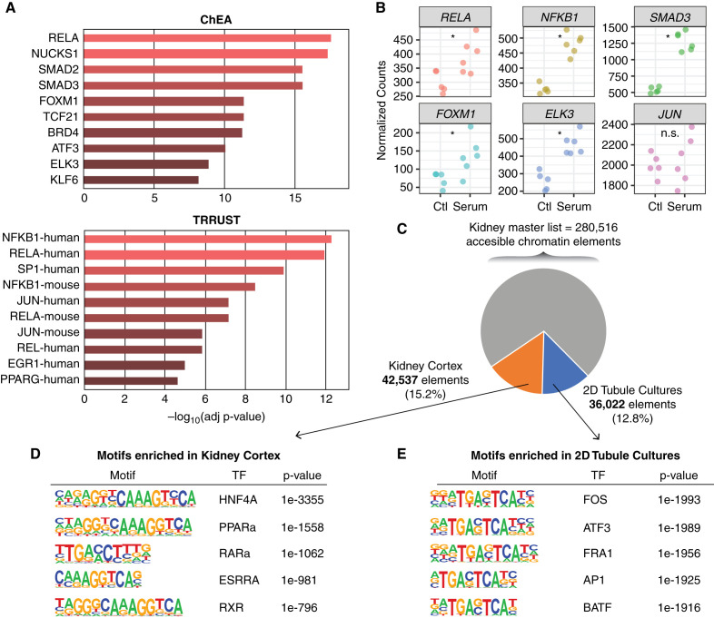
Results: Serum, but not its major protein component albumin, induced tubular injury and secretion of proinflammatory cytokines. Epigenomic comparison of serum-injured tubules and intact kidney tissue revealed canonical stress-inducible regulation of injury-induced genes. Concordant transcriptional changes in microdissected tubulointerstitium were also observed in an independent cohort of patients with proteinuric kidney disease.

Conclusions: Our results demonstrate a causal role for serum proteins in tubular injury and identify regulatory mechanisms and novel pathways for intervention.

Keywords: acute renal failure; albuminuria; blood proteins; gene expression; gene transcription; nephrotic syndrome; proteinuria; proximal tubule; renal tubular epithelial cells; transcription regulation.


Figures

None
Graphical abstract
Figure 1.
Figure 1.
Human kidney tubules in 3D microphysiologic devices exhibit different responses to serum versus albumin exposure. (A) Schematic of two-channel Nortis MPS. The red areas represent a 3D matrix chamber through which a central tubule monolayer–lined channel can be perfused with medium (blue). (B) Compared with control, MPS tubules treated with 2% human serum for 48 hours have a higher frequency of nuclei that label with the proliferative marker Ki-67. (C) Quantification of percentage of Ki-67–positive nuclei in n=5 control and n=4 serum-treated tubular MPSs from donors 1 and 2; P<0.0001 by two-tailed t test. (D) Volcano plot of differentially expressed genes (>1.1× absolute fold-change and adjusted P<0.05) in MPS cultured PTECs treated with 720 µg/ml albumin or 2% human serum for 8 hours. (E) Overlapping significantly differentially expressed genes for serum- and albumin-treated tubule MPSs after 7 days of exposure. A heatmap of 221 shared genes is shown for each donor (n=2–3 per treatment condition) and replicate (n=3 per donor and treatment condition).
Figure 2.
Figure 2.
Serum exposure induces inflammatory cytokine production in kidney tubular epithelial cells. (A) Volcano plot of differentially expressed genes in tubule MPS exposed for 48 hours to 2% human serum versus untreated control. Upregulated, higher expression in serum-treated MPS. Data shown are from two donors with 2–3 replicates per treatment condition. (B) Advaita pathway analysis demonstrates significant perturbations of cytokine, chemokine, TNF, and NF-κB signaling pathway components and ECM-receptor interactions in serum-exposed tubule cells. Dots representing pathways are positioned by their FDR from an effect analysis measuring total perturbation accumulation (pAcc) versus a classic over-representation analysis (pORA). Pathways with FDR<0.05 by both analyses are shown in red. Selected pathways are highlighted in yellow. (C) Individual genes contributing to the enrichment of the top-ranked cytokine-cytokine receptor pathway are shown. (D) Meta-analysis of shared significantly changed pathways (FDR<0.05) between serum-exposed tubule MPSs and primary tubular epithelial cells cultured in 2D in 10% FBS versus intact kidney cortex.
Figure 3.
Figure 3.
Meta-analysis identifies transcriptional and pathway changes induced in serum-exposed tubule MPS that are shared with other preclinical models. (A) Shared differentially expressed genes (DEGs, adjusted P<0.05) among serum-exposed tubule MPS, plasma-treated PTEC cultures grown in 2D, and 3D RPTEC-TERT MPSs injured by cessation of flow. (B) Heatmap of shared DEGs from (A). (C) Shared DEGs among serum-exposed tubule MPSs, a mouse model of Alport syndrome, and a UUO mouse model. (D) Heatmap of shared DEGs from (A). (E) Meta-analysis of shared significantly changed pathways (FDR<0.05) among serum-exposed tubule MPSs, plasma-treated PTEC cultures grown in 2D, and 3D RPTEC-TERT MPSs injured by cessation of flow. (F) Advaita pathway analysis with dots representing pathways positioned by their FDR from an effect analysis measuring total perturbation accumulation (pAcc) versus a classic over-representation analysis (pORA). Yellow dots indicate shared significant pathways from (E) (cytokine-cytokine receptor interaction, viral protein interaction with cytokine-cytokine receptors, and rheumatoid arthritis). All other pathways with FDR<0.05 are shown in red. (G) Meta-analysis of shared significantly changed pathways (FDR<0.05) among serum-exposed tubule MPSs, a mouse model of Alport syndrome, and a UUO mouse model. (F) Advaita pathway analysis with yellow dots denoting shared significant pathways from (G) (NFkB signaling, ECM-receptor interaction, malaria, amoebiasis, complement and coagulation -cascades, and metabolic pathways).
Figure 4.
Figure 4.
AP-1 and NFkB family members are key drivers of the transcriptional landscape of serum-exposed tubules. (A) ENRICHR analysis of serum-exposed tubule MPS DEGs using ChEA and TRRUST libraries, indicating transcription factors (TFs) whose experimentally validated targets are enriched. *Adjusted P<0.05; NS, not significant. (B) Gene expression changes of key TFs implicated by ENRICHR analysis in 3D tubule MPSs exposed to serum. (C) Intact kidney cortex and primary tubules cultured in 2D in 10% FBS share most of their accessible chromatin regions, but also have regions that are only accessible in one sample type. HOMER analysis identifies the top five enriched TF binding motifs in chromatin regions that are more accessible in (D) intact kidney cortex and (E) primary tubules cultured in 2D in 10% FBS.
Figure 5.
Figure 5.
Integrative analysis reveals the regulatory circuitry of HAVCR1. (A) Overview of the HAVCR1 gene locus with gene expression (top) and accessible chromatin (middle) tracks for cultured tubules and intact renal cortex (three donors each). The HAVCR1 gene is expressed at higher levels in tubules cultured in 10% FBS. Green vertical bars indicate regulatory elements with greater accessibility in intact cortex, whereas orange vertical bars indicate regulatory elements with greater accessibility in cultured tubules. The lower panel shows the activity of the same loci in the 733-cell ENCODE regulatory index with elements showing kidney-selective activity highlighted. (B) Comparison of the predicted transcription factor motif archetypes with actual footprints (FDR≤0.001) in tubule DNase-seq data reveals overlap of predicted motifs for AP-1 and NR families in kidney-selective regulatory elements identified in (A).
Figure 6.
Figure 6.
Serum exposure induces secretion of cytokines, matrix metalloproteinases, and shedding of HAVCR1 in 3D MPSs. (A) The log-transformed CPM expression levels of selected genes in the microdissected tubulointerstitium of patients with FSGS (n=79) and MCD (n=38) from the NEPTUNE consortium. All P<0.05 by one-tailed t test. (B) Effluents from serum- and albumin-treated 3D MPSs from three donors were assayed for the indicated proteins by ELISA. All comparisons included at least four replicates for each timepoint and treatment, except for donor 9, for which only one replicate was available for the day 2 and day 4 timepoints. Except where indicated as not significant (NS), all comparisons between serum and control samples were significant at an adjusted P<0.05 using an unpaired t test with Sidak–Holm correction for multiple comparisons. All comparisons between albumin and control were not significant, except for MMP7 for donors 9 and 10 and for KIM-1 for donor 9, which were significant at all timepoints at an adjusted P<0.05 using an unpaired t test with Sidak–Holm correction for multiple comparisons. BLOQ, below limit of quantification.
Figure 7.
Figure 7.
Serum but not albumin exposure induces a proinflammatory response that predisposes to progressive kidney injury and fibrosis.

Comment in

  • Authors' Reply: Serum Protein-induced Tubular Injury.
    Lidberg K, Himmelfarb J, Kelly E, Akilesh S. Lidberg K, et al. J Am Soc Nephrol. 2022 Aug;33(8):1627-1628. doi: 10.1681/ASN.2022060657. J Am Soc Nephrol. 2022. PMID: 35906085 Free PMC article. No abstract available.
  • Serum Protein-Induced Tubular Injury.
    O'Neill WC. O'Neill WC. J Am Soc Nephrol. 2022 Aug;33(8):1627. doi: 10.1681/ASN.2022050568. J Am Soc Nephrol. 2022. PMID: 35906090 Free PMC article. No abstract available.

References

    1. United States Renal Data System NIoH, National Institute of Diabetes and Digestive and Kidney Diseases : 2020 USRDS Annual Data Report: epidemiology of kidney disease in the United States, 2020. Available at: https://adr.usrds.org/2020. Accessed May 1, 2021
    1. Hannedouche T, Landais P, Goldfarb B, el Esper N, Fournier A, Godin M, et al. : Randomised controlled trial of enalapril and beta blockers in non-diabetic chronic renal failure. BMJ 309: 833–837, 1994 - PMC - PubMed
    1. Group MoDiRDS : Effects of dietary protein restriction on the progression of moderate renal disease in the Modification of Diet in Renal Disease Study. J Am Soc Nephrol 7: 2616–2626, 1996 - PubMed
    1. Jafar TH, Stark PC, Schmid CH, Landa M, Maschio G, Marcantoni C, et al. ; AIPRD Study Group. Angiotensin-Converting Enzymne Inhibition and Progression of Renal Disease : Proteinuria as a modifiable risk factor for the progression of non-diabetic renal disease. Kidney Int 60: 1131–1140, 2001 - PubMed
    1. Bakris GL: Slowing nephropathy progression: focus on proteinuria reduction. Clin J Am Soc Nephrol 3[Suppl 1]: S3–S10, 2008 - PMC - PubMed

Publication types