Review
. 2022 Feb 23;10(1):17.
doi: 10.1038/s41413-021-00180-y.

Recent advances in smart stimuli-responsive biomaterials for bone therapeutics and regeneration

Affiliations
Review

Recent advances in smart stimuli-responsive biomaterials for bone therapeutics and regeneration

Hongpu Wei et al. Bone Res. .

Abstract

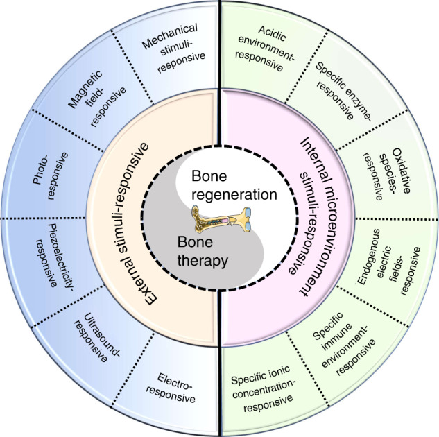
Bone defects combined with tumors, infections, or other bone diseases are challenging in clinical practice. Autologous and allogeneic grafts are two main traditional remedies, but they can cause a series of complications. To address this problem, researchers have constructed various implantable biomaterials. However, the original pathological microenvironment of bone defects, such as residual tumors, severe infection, or other bone diseases, could further affect bone regeneration. Thus, the rational design of versatile biomaterials with integrated bone therapy and regeneration functions is in great demand. Many strategies have been applied to fabricate smart stimuli-responsive materials for bone therapy and regeneration, with stimuli related to external physical triggers or endogenous disease microenvironments or involving multiple integrated strategies. Typical external physical triggers include light irradiation, electric and magnetic fields, ultrasound, and mechanical stimuli. These stimuli can transform the internal atomic packing arrangements of materials and affect cell fate, thus enhancing bone tissue therapy and regeneration. In addition to the external stimuli-responsive strategy, some specific pathological microenvironments, such as excess reactive oxygen species and mild acidity in tumors, specific pH reduction and enzymes secreted by bacteria in severe infection, and electronegative potential in bone defect sites, could be used as biochemical triggers to activate bone disease therapy and bone regeneration. Herein, we summarize and discuss the rational construction of versatile biomaterials with bone therapeutic and regenerative functions. The specific mechanisms, clinical applications, and existing limitations of the newly designed biomaterials are also clarified.


Conflict of interest statement

The authors declare no competing interests.

Figures

Fig. 1
Fig. 1
Scheme summarizing different strategies in the design and fabrication of versatile biomaterials with both therapeutic and regeneration functions
Fig. 2
Fig. 2
Timeline of some representative studies of the application of smart stimuli-responsive biomaterials in the past half-decade. Figures for years 2016–2020: Image for 2016: reproduced with permission. Copyright 2016, Royal Society of Chemistry; Image for 2017: reproduced with permission. Copyright 2017, Elsevier; Image for 2018: reproduced with permission. Copyright 2018, Elsevier; reproduced with permission. Copyright 2018, American Chemical Society; Image for 2019: reproduced with permission. Copyright 2019, Elsevier; reproduced with permission. Copyright 2019, American Chemical Society; reproduced with permission. Copyright 2019, Wiley-VCH; Image for 2020: reproduced with permission. Copyright 2020, American Chemical Society; reproduced with permission. Copyright 2020, Royal Society of Chemistry
Fig. 3
Fig. 3
nHA/GO/CS scaffolds for both disease therapeutic and bone regeneration. a Scheme to manifest the fabrication of nHA/GO/CS scaffolds with bifunctionalities of both therapeutic and regeneration. b Tumor volume changes after different treatments with time (days). c Quantitative analyses of various proteins with or without NIR exposure after 14 days of osteogenic culture (**P < 0.01, *P < 0.05). Reproduced with permission. Copyright 2020, Elsevier
Fig. 4
Fig. 4
A novel biocompatible PDA/IR820/DAP coating for antibiotic/photodynamic/photothermal triple therapy. a Scheme of the antibacterial mechanism of Ti-PDA-IR820-DAP: the synergistical therapy of DAP, PTT, and PDT cause remarkable lethal effect to bacteria. b The results of spread plate assays to show the antibacterial efficiency. c Quantitative analysis of new bone area. d The percentages of bone-implant contact (BIC) were calculated from Van Gieson staining. Reproduced with permission. Copyright 2020, Elsevier
Fig. 5
Fig. 5
3D-printing scaffolds coloading with Fe3O4 and CaO2 NPs (AKT-Fe3O4-CaO2) for cancer therapeutic and bone regeneration. a Scheme of the fabrication of 3DP AKT-Fe3O4-CaO2 scaffold with bifunction of magnetic hyperthermia and bone regeneration. b Time-dependent tumor-volume changes of mice after different treatment (*P < 0.05). c Quantitative analysis of newborn bone tissues after VG stained. d 3D reconstruction of micro-CT images showing the in vivo osteogenesis performance directly (red, newborn bone tissues; white, residual scaffolds). Reproduced with permission. Copyright 2020, Wiley-VCH
Fig. 6
Fig. 6
Rapid photo-sonotherapy for clinical treatment of bacterial infected bone implants by creating oxygen deficiency using sulfur doping. a Scheme of the fabrication of sulfur-doped om titanium implant (Ti-S-TiO2–x), which can enhance sonocatalytic-photothermal ability and manifest exhibits efficient bone infection therapy. b In vitro antibacterial efficiency of 2 kinds of titanium implant in four different conditions for S. aureus from spread plate. c Bacteria colony images to reveal the in vivo antibacterial performance. d The results of the spread plate to reveal the in vivo antibacterial efficiency. e Corresponding calculated new bone area to reveal the in vivo osteogenic performance. f The results of bone volume/total volume (BV/TV) based on the micro-CT results (*P < 0.05, **P < 0.01, ***P < 0.001). Reproduced with permission. Copyright 2020, American Chemical Society
Fig. 7
Fig. 7
A mussel-inspired persistent ROS-scavenging, electroactive, and osteoinductive scaffold based on electrochemical-driven in situ nanoassembly. a Scheme of the fabrication of PPy-PDA-HA-coated scaffolds by curling after coating a PPy-PDA-HA film on a titanium mesh. b Fabrication of the PPy-PDA-HA film by layer-by-layer pulse electrodeposition (LBL-PED) method. c proliferation, and d differentiation of BMSCs on the PPy, PPy-PDA, and PPy-PDA-HA films under different electrical stimulation potentials. e Comparison of bone area (BA) from histomorphometry. Reproduced with permission (*P < 0.05). Copyright 2019, Wiley-VCH
Fig. 8
Fig. 8
3D printing of high-strength bioscaffolds for the synergistic treatment of bone cancer. a Scheme of the synthesis of Fe-CaSiO3 composite scaffolds and their biomedical application. b Changes of the relative tumor volume in the six groups. c Photos of the tumors in six different groups on day 15. d The histomorphometric measurements of the in vivo new bone area in three groups at 8 weeks post-surgery (*P < 0.05). Reproduced with permission. Copyright 2018, Nature Publishing Group
Fig. 9
Fig. 9
Bacteria-triggered pH-responsive osteopotentiating coating on 3D-printed polyetheretherketone scaffolds for infective bone defect repair. a Scheme of the fabrication of 3P-AP-Ag coatings with pH-triggered osteopotentiating properties on 3DP porous PEEK scaffolds. b Schematic of the tests in vitro and in vivo for their multifunction. c Schematic diagram of possible antibacterial factors. Three major factors might contribute to its antibacterial properties: ROS overproduction, Ag+ ion liberation, and surface nanostructure. d Delivery profiles of Ag+ ions from different scaffolds in various pH values (pH = 7.4, 5.0, 4.5). e Delivery profiles of Ca2+ ions from different scaffolds in various pH values (pH = 7.4, 5.0, 4.5). f The bactericidal curves to reveal the antibacterial activities of the coatings. g Quantitative reverse transcription polymerase chain reaction (RT–qPCR) analysis of the gene expressions relates to osteogenesis (ALP, Runx2, Col1a1, and OCN) (*P < 0.05, &P < 0.05). Reproduced with permission. Copyright 2020, American Chemical Society
Fig. 10
Fig. 10
Built‐in electric fields dramatically induce enhancement of osseointegration and bone defect regeneration. a Illustration showing the built-in electric fields promote implant osseointegration: A built-in electric field is constructed among the endogenous electronegative bone defect wall and the electropositive ferroelectric BiFeO3 (BFO) nanofilm implant surface. Thus, the rapid and high-quality osseointegration was induced on the implant. b Schematic illustration of the built-in electric fields forming among BFO+ nanofilm implants and bone. And the corresponding histological analysis at 2 weeks post-implantation, which showing better osseointegration on BFO+ nanofilm implants (NB: nascent bone). c Diagram of quantitative analysis of bone-implant contact (BIC) values. d Diagram of different bone volume/total volume (BV/TV) on the basis of histomorphometry analysis (*P < 0.05, **P < 0.01). e Graphical summary of phases of BFO+ nanofilm-trigged osteointegration. Reproduced with permission. Copyright 2017, Wiley-VCH
Fig. 11
Fig. 11
Systemic administration of enzyme-responsive growth factor nanocapsules for promoting bone repair. a Illustration showing the mechanism of enzyme-responsive BMP-2 nanocapsule (n(BMP-2)) and their responsive delivery for bone fracture repair. b The distribution of BMP-2 and n(BMP-2) in bone defect site and other tissues site after intravenous injection of BMP-2 and n(BMP-2). c Comparation of the expression of alkaline phosphatase (ALP) activity of MSCs with BMP-2 or n(BMP-2) incubated (****P < 0.000 1). d Comparation of the Micro-CT images of different therapy after rat tibial fracture. e Comparation of the volume of bone tissue per volume of total tissue (BV/TV) after different therapy (**P < 0.01, ***P < 0.001). Reproduced with permission. Copyright 2019, Royal Society of Chemistry
Fig. 12
Fig. 12
Summative scheme of the current research developments and the future outlook in smart stimuli-responsive biomaterials with multiple functions of bone therapeutics and bone regeneration

Similar articles

Cited by

References

    1. Zhang Y, et al. Advancements in hydrogel-based drug sustained release systems for bone tissue engineering. Front. Pharmacol. 2020;11:622. - PMC - PubMed
    1. Wang P, et al. Bone tissue engineering via nanostructured calcium phosphate biomaterials and stem cells. Bone Res. 2014;2:14017. - PMC - PubMed
    1. Zhang K, et al. Advanced smart biomaterials and constructs for hard tissue engineering and regeneration. Bone Res. 2018;6:31. - PMC - PubMed
    1. Dang M, Saunders L, Niu X, Fan Y, Ma PX. Biomimetic delivery of signals for bone tissue engineering. Bone Res. 2018;6:25. - PMC - PubMed
    1. Sun W, et al. Injectable nano-structured silicon-containing hydroxyapatite microspheres with enhanced osteogenic differentiation and angiogenic factor expression. Ceram. Int. 2018;44:20457–20464.