. 2022 Feb 23;7(1):24.
doi: 10.1038/s41541-022-00450-8.

An adjuvanted subunit SARS-CoV-2 spike protein vaccine provides protection against Covid-19 infection and transmission

Affiliations

An adjuvanted subunit SARS-CoV-2 spike protein vaccine provides protection against Covid-19 infection and transmission

Kairat Tabynov et al. NPJ Vaccines. .

Abstract

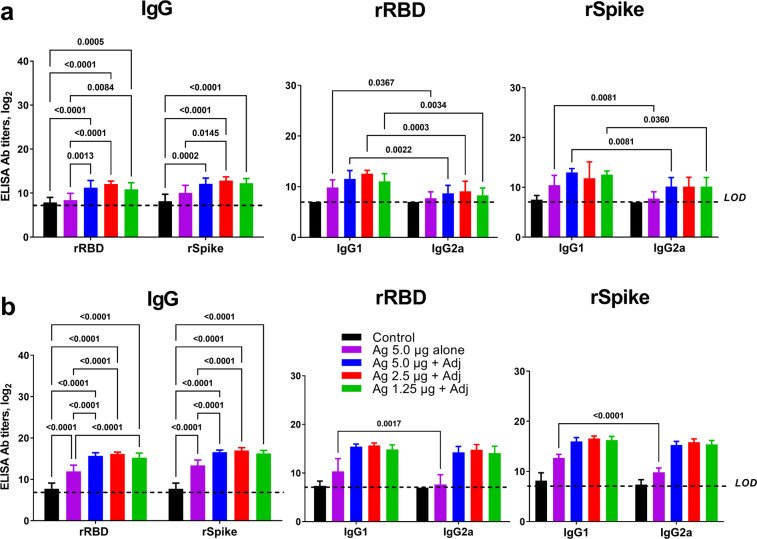
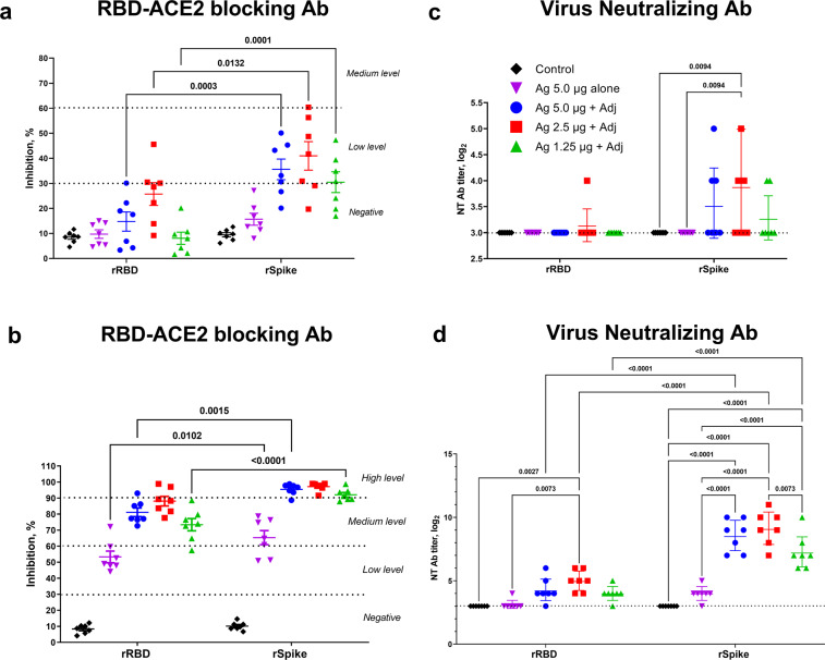
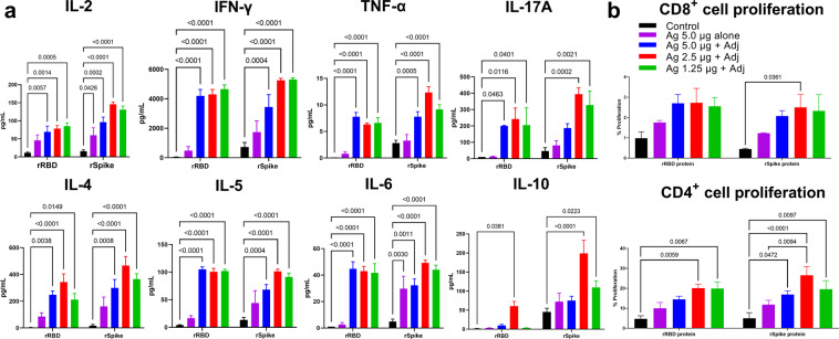
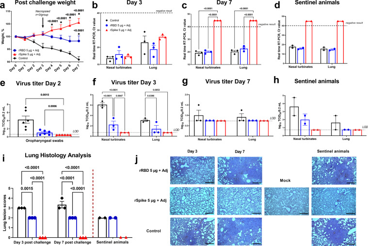
Recombinant protein approaches offer major promise for safe and effective vaccine prevention of SARS-CoV-2 infection. We developed a recombinant spike protein vaccine (called NARUVAX-C19) and characterized its ability when formulated with a nanoemulsion adjuvant to induce anti-spike antibody and T-cell responses and provide protection including against viral transmission in rodent. In mice, NARUVAX-C19 vaccine administered intramuscularly twice at 21-day interval elicited balanced Th1/Th2 humoral and T-cell responses with high titers of neutralizing antibodies against wild-type (D614G) and delta (B.1.617.2) variants. In Syrian hamsters, NARUVAX-C19 provided complete protection against wild-type (D614G) infection and prevented its transmission to naïve animals (n = 2/group) placed in the same cage as challenged animals (n = 6/group). The results contrasted with only weak protection seen with a monomeric spike receptor-binding domain (RBD) vaccine even when formulated with the same adjuvant. These encouraging results warrant the ongoing development of this COVID-19 vaccine candidate.


Conflict of interest statement

NP and LL are affiliated with Vaxine Pty Ltd which holds the rights to COVAX-19 vaccine. None of the other authors has any financial or personal interest with any organization that could inappropriately influence or bias the research activity presented in this manuscript.

Figures

Fig. 1
Fig. 1. Antigen-specific IgG, IgG1 and IgG2a titers in serum of BALB/c mice vaccinated with rRBD or rSpike protein ECD at 21 days after prime and booster immunization.
a Data post prime vaccination, b data post booster vaccination. Antigens were used at doses of 1.25, 2.5, and 5 µg with SWE adjuvant. For comparison, 5 µg vaccine was given without adjuvant and a negative control vaccine comprised SWE adjuvant with no antigen. Antibody levels are presented as geometric mean titers (GMT) with a 95% confidence interval. Differences in IgG titers were assessed using Tukey’s multiple comparisons test. A P < 0.05 value was considered as a significant difference.
Fig. 2
Fig. 2. RBD-ACE2 blocking and virus-neutralizing antibody titers in BALB/c mice at 21 days after prime and booster immunization with SWE-adjuvanted rRBD and rSpike protein vaccines.
a, c Data post prime vaccination, b, d data post booster vaccination. Controls included rRBD or rSpike protein alone (Ag 5.0 µg alone) or SWE adjuvant alone (Control). RBD-ACE2 blocking antibodies were determined according to the level of inhibition: negative (<30%), low (30–59%), medium (60–89), and high (90≤). Virus neutralizing antibody levels are presented as geometric mean titers (GMT) with a 95% confidence interval. Differences in antibody titers between groups were assessed using Tukey’s multiple comparisons test.
Fig. 3
Fig. 3. Antigen-stimulated cytokine production and CD4 + and CD8 + T cell proliferation in splenocytes from rRBD or rSpike protein vaccinated BALB/c mice.
Controls included rRBD or rSpike protein alone at 5.0 µg (Ag 5.0 µg alone) and SWE adjuvant-alone group (Control). Cytokine data (a) were presented as the difference (delta) in cytokine concentrations between samples with and without protein stimulation. CD4 + and CD8 + T cell proliferation (b) was calculated as the difference (Δ) in number of proliferating (CFSE + ) lymphocytes between stimulated vs non-stimulated cells. Statistical differences were assessed using Šídák’s multiple comparisons test. A P < 0.05 value was considered as a significant difference.
Fig. 4
Fig. 4. Spike-specific IgG, RBD-ACE2 blocking antibody and neutralizing antibody levels in hamsters at 21 days after booster immunization with SWE-adjuvanted rRBD or rSpike protein as compared to convalescent sera.
a Data for spike-specific IgG, b RBD-ACE2 blocking antibody, c, d neutralizing antibody levels. Control refers to a group of animals injected with adjuvant-alone with no antigen. Convalescent refers to post-infection serum samples from four unvaccinated hamsters that had previously been challenged with the wild-type SARS-CoV-2 virus. Viral neutralizing antibodies were assessed against wild-type D614G (c) and delta variant (d) viruses. Differences between groups assessed using Dunnett’s multiple comparisons test.
Fig. 5
Fig. 5. Vaccine protection in Syrian hamsters against SARS-CoV-2 infection and virus transmission.
To assess vaccine efficacy animals (n = 6/group) were intranasally infected with WT SARS-CoV-2 and the following parameters then measured: 1) changes in body weight (a; n = 3-6/group); virus excretion from oropharyngeal swabs Day 2 post challenge (e; n = 6/group) 2) viral load in nasal turbinates (n = 3/group) and lungs (n = 3/group) by PCR (represented by Ct; b, c) and culture of infectious virus (expressed as log10 TCID50/0.2 mL; f, g); 3) histopathological changes in the lungs (i, j), on Days 3 (n = 3/group) and 7 (n = 3/group) after challenge. Efficacy against virus transmission in contact sentinel animals (n = 2/group) was evaluated by detection of viral load (d, h), lung pathology scoring (i, j). Control included an adjuvant-alone group. Scale bars are 500 µm. Differences in the studied parameters between animal groups were assessed using Tukey’s (virus titer; Lung Histology), Dunnett’s (postchallenge weight), or Šídák’s (viral load in PCR) multiple comparisons test. A P < 0.05 value was considered as a significant difference. * in comparison with the control group. LOD limit of detection.
Fig. 6
Fig. 6. Correlation matrix analysis between immune markers including various antibody measurements and measures of vaccine protection including weight changes, viral loads, and lung pathology.
The color refers to r value scale (−1 to 1) shown on the right. The number in each cell indicates the actual r value and the lower digits represent p-values. The analysis was conducted by multivariable Pearson correlation method.

References

    1. World Health Organization. Coronavirus disease (COVID-19) pandemic. 2021. [accessed 2021 Nov]. https://www.who.int/emergencies/diseases/novel-coronavirus-2019.
    1. Planas D, et al. Reduced sensitivity of SARS-CoV-2 variant Delta to antibody neutralization. Nature. 2021;596:276–280. doi: 10.1038/s41586-021-03777-9. - DOI - PubMed
    1. Weisblum Y, et al. Escape from neutralizing antibodies by SARS-CoV-2 spike protein variants. Elife. 2020;9:e61312. doi: 10.7554/eLife.61312. - DOI - PMC - PubMed
    1. Singanayagam A, et al. Community transmission and viral load kinetics of the SARS-CoV-2 delta (B.1.617.2) variant in vaccinated and unvaccinated individuals in the UK: a prospective, longitudinal, cohort study. Lancet Infect. Dis. 2021;S1473-3099:00648–4. - PMC - PubMed
    1. World Health Organization. COVID-19 vaccine tracker and landscape. [accessed 2021 Aug 03]. https://www.who.int/publications/m/item/draft-landscape-of-covid-19-cand....