. 2022 Feb 23;12(1):3056.
doi: 10.1038/s41598-022-06848-7.

Endothelial cell ferroptosis mediates monocrotaline-induced pulmonary hypertension in rats by modulating NLRP3 inflammasome activation

Affiliations

Endothelial cell ferroptosis mediates monocrotaline-induced pulmonary hypertension in rats by modulating NLRP3 inflammasome activation

Shan-Shan Xie et al. Sci Rep. .

Abstract

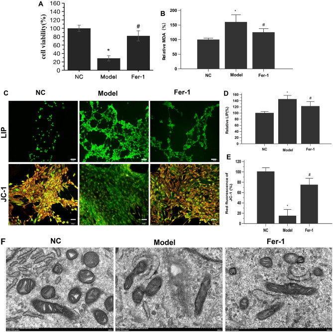
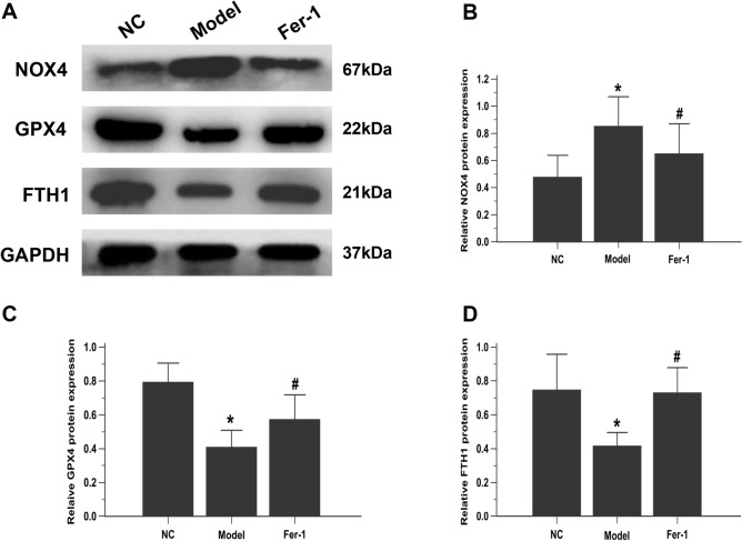
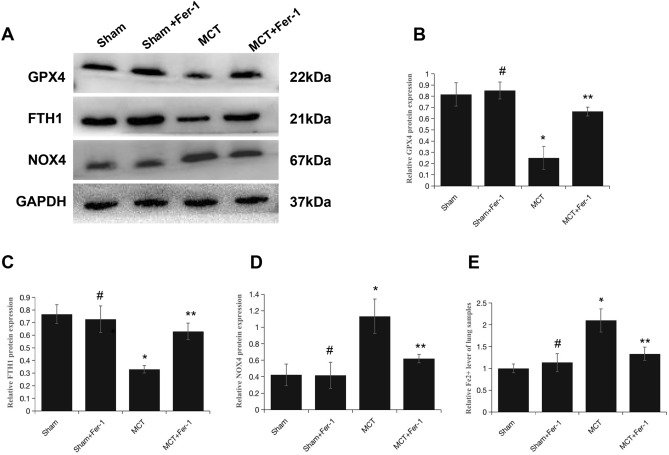
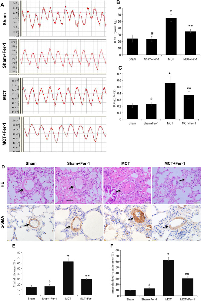
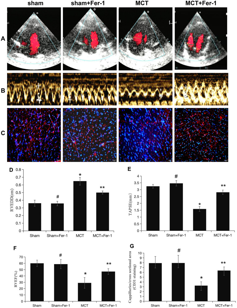
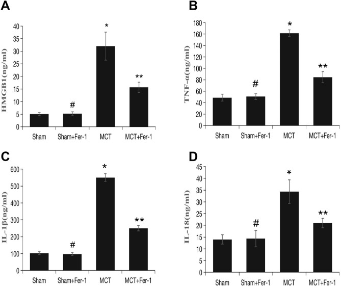
Inflammation triggers pulmonary vascular remodelling. Ferroptosis, a nonapoptotic form of cell death that is triggered by iron-dependent lipid peroxidation and contributes to the pathogenesis of several inflammation-related diseases, but its role in pulmonary hypertension (PH) has not been studied. We examined endothelial cell ferroptosis in PH and the potential mechanisms. Pulmonary artery endothelial cells (PAECs) and lung tissues from monocrotaline (MCT)-induced PH rats were analysed for ferroptosis markers, including lipid peroxidation, the labile iron pool (LIP) and the protein expression of glutathione peroxidase 4 (GPX4), ferritin heavy chain 1 (FTH1) and NADPH oxidase-4 (NOX4). The effects of the ferroptosis inhibitor ferrostatin-1 (Fer-1) on endothelial cell ferroptosis and pulmonary vascular remodelling in MCT-induced rats were studied in vitro and in vivo. Ferroptosis was observed in PAECs from MCT-induced PH rats in vitro and in vivo and was characterized by a decline in cell viability accompanied by increases in the LIP and lipid peroxidation, the downregulation of GPX4 and FTH1 expression and the upregulation of NOX4 expression. High-mobility group box 1 (HMGB1)/Toll-like receptor 4 (TLR4)/NOD-like receptor family pyrin domain containing 3 (NLRP3) inflammasome signalling was measured by western blotting. These changes were significantly blocked by Fer-1 administration in vitro and in vivo. These results suggest that Fer-1 plays a role in inhibiting ferroptosis-mediated PAEC loss during the progression of PH. The ferroptosis-induced inflammatory response depended on the activation of HMGB1/TLR4 signalling, which activated the NLRP3 inflammasome in vivo. We are the first to suggest that pulmonary artery endothelial ferroptosis triggers inflammatory responses via the HMGB1/TLR4/NLRP3 inflammasome signalling pathway in MCT-induced rats. Treating PH with a ferroptosis inhibitor and exploring new treatments based on ferroptosis regulation might be promising therapeutic strategies for PH.


Conflict of interest statement

The authors declare no competing interests.

Figures

Figure 1
Figure 1
Ferroptosis occurs in PAECs derived from MCT-induced PH rats. (A) Fer-1 restored the viability of MCT-induced PH rat-derived PAECs. (B) Fer-1 reduced lipid peroxidation in MCT-induced PH rat-derived PAECs. (C) Representative LIP and JC-1-stained PAECs. Morphology was examined using light microscopy. Scale bar = 100 μm. (D) Quantification of the relative LIP value in the different groups of PAECs. (E) Quantification of the relative value of JC-1 red fluorescence in the different groups of PAECs. (F) Representative TEM images of PAECs in the different groups show mitochondrial morphology. PAECs in the model group showed mitochondrial damage with mitochondrial outer membrane rupture and mitochondrial crista disappearance. Fer-1 restored these changes. Scale bar = 500 nm. NC, normal control group; Model, model group; Fer-1, MCT + Fer-1 (10 nM, treated for 4 h) group. Each experiment was repeated 3 times, n = 3. The data represent the means ± SD. *P < 0.05 versus the NC group, #P < 0.05 versus the model group.
Figure 2
Figure 2
Fer-1 downregulates ferroptosis markers in PAECs. (A) Representative images showing the effect of Fer-1 on the expression of ferroptosis-related proteins (NOX4, GPX4 and FTH1) in PAECs, as assessed by western blot analysis. The group of blots were cropped from the same gel. (BD) Bar graph showing that Fer-1 treatment significantly restored ferroptosis-related proteins (NOX4, GPX4 and FTH1) in PAECs. Quantitative western blot results were normalized to GAPDH. NC, normal control group; Model, model group; Fer-1, MCT + Fer-1 (10 nM) group. Each experiment was repeated 3 times, n = 3. The data represent the means ± SD. *P < 0.05 versus the NC group, #P < 0.05 versus the model group.
Figure 3
Figure 3
Fer-1 downregulates ferroptosis markers in MCT-induced PH rats. (A) Representative western blots showing ferroptosis-related proteins (NOX4, GPX4 and FTH1) in total lung homogenates from PH rats 4 weeks after MCT exposure. (BD) The quantification of protein expression is shown; GAPDH was used as a loading control (37 kDa). The groups of blots were cropped from the same gel. (E) Bar graph showing Fer-1-mediated downregulation of the level of Fe2+ in MCT-induced PH rats. The data are shown as the mean ± SD, #P > 0.05 versus the sham group, *P < 0.05 versus the sham group, **P < 0.05 versus the MCT group. n = 8–12/group.
Figure 4
Figure 4
Fer-1 ameliorated MCT-induced haemodynamics, RV hypertrophy and pulmonary vascular remodelling in rats. (A) Representative RV pressure curves in the sham, sham + Fer-1, MCT, and MCT + Fer-1 groups. The MCT + Fer-1 group was pretreated with Fer-1 (2 mg/kg/d). (B) Bar graph comparing RVSPs in the different groups of rats. (C) Fer-1 pretreatment reduced the RV/LV + S ratio of MCT-induced rats. (D) Representative image of haematoxylin–eosin (HE) staining showing the expression of α-SMA to compare arteriole wall thickness in the sham, sham + Fer-1, MCT, and MCT + Fer-1 pretreatment groups. (E) Quantification of arteriole percent medial thickness. (F) The percentage of muscularization is shown by staining for α-SMA. The data represent the means ± SD. #P > 0.05 versus the sham group, *P < 0.05 versus the sham group, **P < 0.05 versus the MCT group. n = 8–12/group.
Figure 5
Figure 5
Pretreatment with Fer-1 attenuates RV function and capillaries of the RV in MCT-induced rats. (A, B) Representative colour Doppler images of the four chambers and TAPSE in the right ventricle in the sham, sham + Fer-1, MCT, and MCT + Fer-1 groups. (C) Representative microphotographs of right ventricles that were stained with DAPI (blue) and immunostained for CD31 (red). (D-F) Quantification of RVEDD, TAPSE, and RVEF. (G) Bar graph showing the numbers of capillaries in the right ventricles of the different groups of rats. The values represent the mean ± SD. #P > 0.05 versus the sham group, *P < 0.05 versus the sham group, **P < 0.05 versus the MCT group. n = 8–12/group.
Figure 6
Figure 6
PAEC ferroptosis induces HMGB1 release, upregulates macrophage TLR4 expression and activates the NLRP3 inflammasome in vitro. (A) Bar graph showing that Fer-1 treatment significantly inhibited HMGB1 release in PAECs derived from MCT-induced rats. (B) Representative images showing the effect of Fer-1 on TLR4 expression in cocultured macrophages (NR8383 cell line). (C) Bar graph showing that Fer-1 treatment significantly restored TLR4 protein expression in cocultured macrophages. (D-J) TAK-242 significantly inhibited the increase in TLR4 and restored the upregulated expression of NLRP3 inflammasome-related proteins (ASC, NLRP3, pro-caspase1, and caspase1 and ratio of caspase1/pro-caspase1). The group of blots were cropped from the same gel. (K-M) Effects of TAK-242 on the secretion of TNF-α, IL-1β, and IL-18 in cocultured macrophages. Each experiment was repeated 3 times, n = 3. HMGB1, High-mobility group box-1 protein. *P < 0.05 versus the negative group; #P < 0.05 versus the HMGB1 group.
Figure 7
Figure 7
Fer-1 pretreatment inhibits lung TLR4/NLRP3 inflammasome signalling activation in MCT-induced rats. (A) Representative western blot showing the expression of TLR4 and NLRP3 inflammasome markers (ASC, NLRP3, pro-caspase1, caspase1) in lung samples from the different groups of rats. The groups of blots were cropped from the same gel. (B-G) Bar graph showing Fer-1-mediated downregulation of TLR4 and NLRP3 inflammasome markers (ASC, NLRP3, pro-caspase1, caspase1 and ratio of caspase1/pro-caspase1) expression in MCT-induced PH rats. Relative protein levels were determined after normalization to GAPDH. The data represent the means ± SD. #P > 0.05 versus the sham group, *P < 0.05 versus the sham group, **P < 0.05 versus the MCT group. n = 8–12/group.
Figure 8
Figure 8
Effect of Fer-1 on HMGB1 expression and inflammatory status in rats. (A) Bar graph showing Fer-1-mediated inhibition of HMGB1 expression in the lung. (B-D) Tumour necrosis factor-α (TNF-α), interleukin-1β (IL-1β) and interleukin-18 (IL-18) in lung tissue. The data represent the means ± SD. #P > 0.05 versus the sham group, *P < 0.05 versus the sham group, **P < 0.05 versus the MCT group. n = 8–12/group.

References

    1. Hoeper MM, et al. Pulmonary Hypertension. Dtsch Arztebl Int. 2017;114:73–84. doi: 10.3238/arztebl.2017.0073. - DOI - PMC - PubMed
    1. Guignabert C, et al. New molecular targets of pulmonary vascular remodeling in pulmonary arterial hypertension: importance of endothelial communication. Chest. 2015;147:529–537. doi: 10.1378/chest.14-0862. - DOI - PubMed
    1. Thenappan T, Ormiston ML, Ryan JJ, Archer SL. Pulmonary arterial hypertension: Pathogenesis and clinical management. BMJ. 2018;360:j5492. doi: 10.1136/bmj.j5492. - DOI - PMC - PubMed
    1. Lajoie AC, et al. Combination therapy versus monotherapy for pulmonary arterial hypertension: A meta-analysis. Lancet Respir. Med. 2016;4:291–305. doi: 10.1016/S2213-2600(16)00027-8. - DOI - PubMed
    1. Stockwell BR, et al. Ferroptosis: A regulated cell death nexus linking metabolism, redox biology, and disease. Cell. 2017;171:273–285. doi: 10.1016/j.cell.2017.09.021. - DOI - PMC - PubMed

Publication types

MeSH terms