

Not Only in Sensorimotor Network: Local and Distant Cerebral Inherent Activity of Chronic Ankle Instability-A Resting-State fMRI Study
- PMID: 35197822
- PMCID: PMC8859266
- DOI: 10.3389/fnins.2022.835538
Not Only in Sensorimotor Network: Local and Distant Cerebral Inherent Activity of Chronic Ankle Instability-A Resting-State fMRI Study
Abstract
Background: Increasing evidence has proved that chronic ankle instability (CAI) is highly related to the central nervous system (CNS). However, it is still unclear about the inherent cerebral activity among the CAI patients.
Purpose: To investigate the differences of intrinsic functional cerebral activity between the CAI patients and healthy controls (HCs) and further explore its correlation with clinical measurement in CAI patients.
Materials and methods: A total of 25 CAI patients and 39 HCs were enrolled in this study. Resting-state functional magnetic resonance imaging (rs-fMRI) was used to detect spontaneous cerebral activity. The metrics of amplitude of low-frequency fluctuation (ALFF), fractional ALFF (fALFF), and regional homogeneity (ReHo) of the two groups were compared by two-sample t-test. The brain regions that demonstrated altered functional metrics were selected as the regions of interest (ROIs). The functional connectivity (FC) was analyzed based on the ROIs. The Spearman correlation was calculated between rs-fMRI metrics and clinical scale scores.
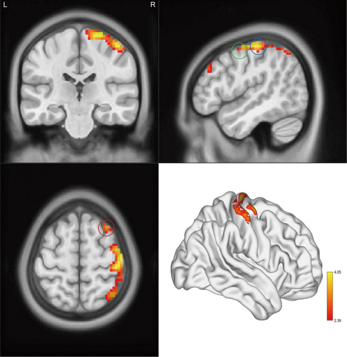
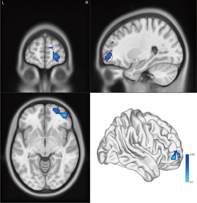
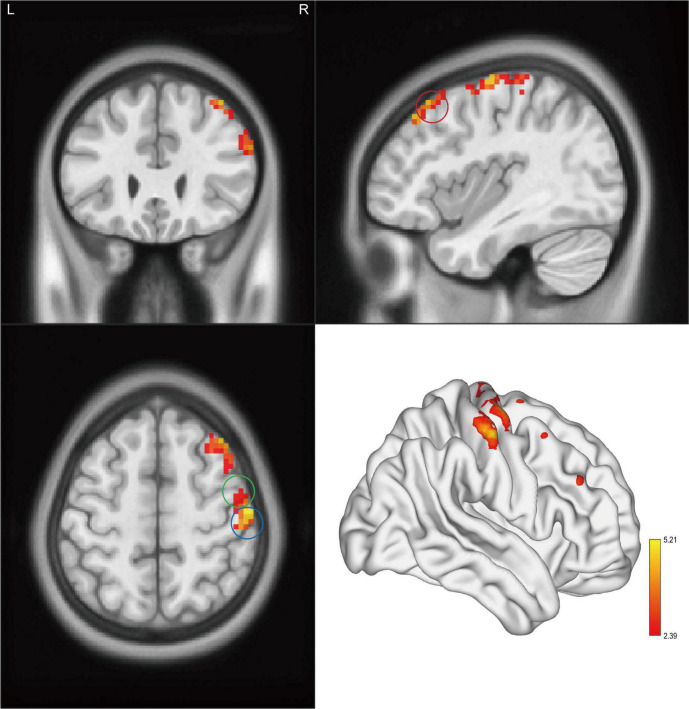
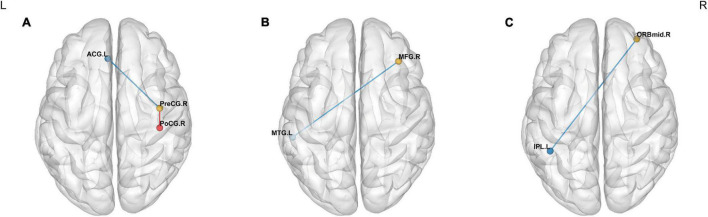
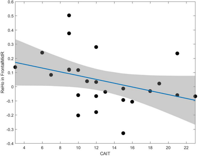
Results: Compared with HCs, CAI patients showed higher ALFF and ReHo values in the right postcentral gyrus, the right precentral gyrus, and the right middle frontal gyrus, while lower fALFF values in the orbital-frontal cortex (OFC, p < 0.01 after correction). Increasing FC between the right precentral gyrus and the right postcentral gyrus while decreasing FC between the right precentral gyrus and the anterior cingulum cortex (ACC), the right middle frontal gyrus and the left middle temporal gyrus, and the OFC and left inferior parietal lobule (IPL) was observed. In addition, in the CAI group, the ReHo value negatively correlated with the Cumberland Ankle Instability Tool score in the right middle frontal gyrus (r = -0.52, p = 0.007).
Conclusion: The CAI patients exhibited enhanced and more coherent regional inherent neuronal activity within the sensorimotor network while lower regional inherent activity in pain/emotion modulation related region. In addition, the information exchanges were stronger within the sensorimotor network while weaker between distant interhemispheric regions. Besides, the increased inherent activity in the right middle frontal gyrus was related to clinical severity. These findings may provide insights into the pathophysiological alteration in CNS among CAI patients.
Keywords: amplitude of low-frequency fluctuation; chronic ankle instability; functional connectivity; regional homogeneity; resting-state fMRI.
Copyright © 2022 Shen, Wang, Wang, Yang, Yuan, Yang, Wu, Wang, Deng, Wang and Liu.
Conflict of interest statement
The authors declare that the research was conducted in the absence of any commercial or financial relationships that could be construed as a potential conflict of interest.
Figures
References
LinkOut - more resources
Full Text Sources