. 2022 Feb 7:12:812627.
doi: 10.3389/fimmu.2021.812627. eCollection 2021.

Increased Fibrosis in a Mouse Model of Anti-Laminin 332 Mucous Membrane Pemphigoid Remains Unaltered by Inhibition of Aldehyde Dehydrogenase

Affiliations

Increased Fibrosis in a Mouse Model of Anti-Laminin 332 Mucous Membrane Pemphigoid Remains Unaltered by Inhibition of Aldehyde Dehydrogenase

Sabrina Patzelt et al. Front Immunol. .

Abstract

Mucous membrane pemphigoid (MMP) is an autoimmune blistering disease characterized by autoantibodies against the basal membrane zone of skin and surface-close epithelia and predominant mucosal lesions. The oral cavity and conjunctivae are most frequently affected, albeit clinical manifestations can also occur on the skin. MMP-associated lesions outside the oral cavity typically lead to scarring. Mechanisms underlying scarring are largely unknown in MMP and effective treatment options are limited. Herein, we assessed the collagen architecture in tissue samples of an antibody-transfer mouse model of anti-laminin-332 MMP. In MMP mice, increased collagen fibril density was observed in skin and conjunctival lesions compared to mice injected with normal rabbit IgG. The extracellular matrix of MMP skin samples also showed altered post-translational collagen cross-linking with increased levels of both lysine- and hydroxylysine-derived collagen crosslinks supporting the fibrotic phenotype in experimental MMP compared to control animals. In addition, we evaluated a potential anti-fibrotic therapy in experimental anti-laminin-332 MMP using disulfiram, an inhibitor of the aldehyde dehydrogenase (ALDH), which has been implicated in immune-mediated mucosal scarring. In addition, disulfiram also acts as a copper chelator that was shown to block lysyl oxidase activity, an enzyme involved in formation of collagen crosslinks. Topical use of disulfiram (300 μM in 2% [w/v] methocel) did not improve ocular lesions in experimental MMP over the 12-day treatment period in disulfiram-treated mice compared to vehicle-treated mice (n=8/group). Furthermore, C57BL6/J mice (n=8/group) were treated prophylactically with 200 mg/kg p.o. disulfiram or the solvent once daily over a period of 12 days. Systemic treatment did not show any reduction in the severity of oral and ocular lesions in MMP mice, albeit some improvement in skin lesions was observed in disulfiram- vs. vehicle-treated mice (p=0.052). No reduction in fibrosis was seen, as assessed by immunohistochemistry. Whilst blocking of ALDH failed to significantly ameliorate disease activity, our data provide new insight into fibrotic processes highlighting changes in the collagenous matrix and cross-linking patterns in IgG-mediated MMP.

Keywords: aldehyde dehydrogenase; autoimmune blistering disease; collagen; crosslinking; fibrosis; laminin 332; mouse model; mucous membrane pemphigoid.


Conflict of interest statement

The authors declare that the research was conducted in the absence of any commercial or financial relationships that could be construed as a potential conflict of interest.

Figures
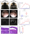
Figure 1
Figure 1
The mouse model of mucous membrane pemphigoid (MMP) recapitulated phenotypic features of human disease up to four weeks. (A) Throughout the 28-day study, injection of anti-laminin alpha 3 (Lama3) IgG led to development of crusts and erosions on the skin mainly presenting on the head and neck area (n=8/group). One mouse/group died during the course of the study. (B) Endoscopy showed prominent oral lesions in MMP mice on Day 28. (C) Anti-Lama3 IgG-injected mice presented with periocular crusted erosions and conjunctival inflammation. (D) Consistent with the presence of ocular lesions, histological analysis showed subepithelial cleavage and leukocyte infiltrates in palpebral conjunctiva biopsies of MMP mice (D). Mice injected with normal rabbit IgG served as a control and did not show any meaningful clinical or histological manifestations. A two-way ANOVA with Sidak’s multiple comparisons test was used to calculate significance (A, C).
Figure 2
Figure 2
Experimental mucous membrane pemphigoid (MMP) showed an increased collagen fibril density. (A, B) MMP mice injected with anti-laminin alpha 3 (Lama3) IgG showed increased collagen density in lesional skin (A) and palpebral conjunctiva (B) on Day 28, as assessed by Picrosirius red staining (top row of panels A and B) and Masson’s trichrome staining (bottom row of panels A and B). No increases in collagen fibril density were seen in control mice treated with normal rabbit IgG. Scale bars = 100 µm.
Figure 3
Figure 3
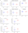
Experimental mucous membrane pemphigoid (MMP) showed elevated levels of collagen crosslinks. (A, B) Amino acid analysis showed no changes in collagen content (A) as well as non-collagenous proteins (B) in MMP vs. control mice. (C) Significant increases in total collagen cross-links as well as HLNL were observed in MMP lesional skin samples, compared with healthy control mice. In addition, higher levels of DHLNL were measured in MMP lesional skin, whereas levels of the mature cross-link HP were comparable in both groups. HP, Hydroxyllysylpyridinoline; DHLNL, dihydroxylysinonorleucine; HLNL, hydroxylysinonorleucine.
Figure 4
Figure 4
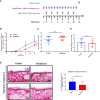
Local use of the ALDH inhibitor disulfiram did not improve conjunctival lesions in experimental MMP. (A) Disulfiram or the respective solvent were administered to the diseased mice (n = 8/group) daily from Day 8 to Day 20 as eye drops. (B) Mice with experimental MMP presented with progressive conjunctival scarring and corneal changes on Day 20. Statistical analysis was performed using two-way ANOVA with Sidak’s multiple comparisons test. (C) On Day 8, the mice had an average affected ocular surface area of approximately 20%. Over the treatment period of 12 days, no improvement in the extent of ocular lesions was observed as a result of the topical treatment in both groups. (D) No significant difference in conjunctiva scores was present in vehicle- vs. disulfiram-treated tissue biopsies. n.s., not significant.
Figure 5
Figure 5
Systemic treatment with the ALDH inhibitor disulfiram did not resolve skin and mucosal lesions in experimental MMP. (A) C57BL6/J mice (n = 8/group) were treated prophylactically with p.o. 200 mg/kg disulfiram once daily or the respective solvent, i.e. corn oil, over a period of 12 days. (B) No significant changes in affected body surface area were observed in both treatment groups, though disulfiram-treated mice showed a trend towards improvement in clinical skin scores (p = 0.0521). Statistical analysis was performed using two-way ANOVA with Tukey’s multiple comparisons test. (C, D) In addition, endoscopy and histological analysis showed no improvement in the severity of oral mucosal lesions (C) or conjunctiva scores (D), respectively, in vehicle- vs. disulfiram-treated mice. (E) No significant reduction in collagen fibril density was seen in vehicle vs. disulfiram-treated mice, as assessed by Picrosirius red staining. Scale bar = 50 µm. n.s., not significant; SD, standard deviation.

References

    1. Rashid H, Lamberts A, Borradori L, Alberti-Violetti S, Barry RJ, Caproni M, et al. . European Guidelines (S3) on Diagnosis and Management of Mucous Membrane Pemphigoid, Initiated by the European Academy of Dermatology and Venereology - Part I. J Eur Acad Dermatol Venereol (2021) 35:1750–64. doi: 10.1111/jdv.17397 - DOI - PMC - PubMed
    1. Schmidt E, Rashid H, Marzano AV, Lamberts A, Di Zenzo G, Diercks GFH, et al. . European Guidelines (S3) on Diagnosis and Management of Mucous Membrane Pemphigoid, Initiated by the European Academy of Dermatology and Venereology - Part II. J Eur Acad Dermatol Venereol (2021) 35:1926–48. doi: 10.1111/jdv.17395 - DOI - PMC - PubMed
    1. Schmidt E, Zillikens D. Pemphigoid Diseases. Lancet (2013) 381:320–32. doi: 10.1016/S0140-6736(12)61140-4 - DOI - PubMed
    1. Kamaguchi M, Iwata H. The Diagnosis and Blistering Mechanisms of Mucous Membrane Pemphigoid. Front Immunol (2019) 10:34. doi: 10.3389/fimmu.2019.00034 - DOI - PMC - PubMed
    1. Georgoudis P, Sabatino F, Szentmary N, Palioura S, Fodor E, Hamada S, et al. . Ocular Mucous Membrane Pemphigoid: Current State of Pathophysiology, Diagnostics and Treatment. Ophthalmol Ther (2019) 8:5–17. doi: 10.1007/s40123-019-0164-z - DOI - PMC - PubMed

Publication types