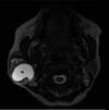
Preoperative Challenges in Managing Intraparotid Schwannoma
- PMID: 35198300
- PMCID: PMC8856645
- DOI: 10.7759/cureus.21392
Preoperative Challenges in Managing Intraparotid Schwannoma
Abstract
Schwannomas are a benign and rare entity that originates from Schwann cells. The majority of schwannomas are found in the head and neck regions and usually involve the intratemporal course of the facial nerve (FN). Isolated extratemporal intraparotid involvement is very rare. It is very challenging to diagnose intraparotid facial nerve schwannoma (PFNS) based on fine needle aspiration cytology (FNAC) preoperatively. We report a case of an intraparotid facial nerve schwannoma masquerading as pleomorphic adenoma. The diagnostic challenges and imaging features along with its management are discussed.
Keywords: facial nerve; head and neck neoplasms; imaging; parotid; schwannoma.
Copyright © 2022, Sultan Abdul Kader et al.
Conflict of interest statement
The authors have declared that no competing interests exist.
Figures


References
-
- Intraparotid facial nerve schwannoma: a report of five cases and an analysis of MR imaging results. Shimizu K, Iwai H, Ikeda K, Sakaida N, Sawada S. http://www.ajnr.org/content/26/6/1328.long. AJNR Am J Neuroradiol. 2005;26:1328–1330. - PMC - PubMed
-
- The intraparotid facial nerve schwannoma: a diagnostic and management conundrum. Gross BC, Carlson ML, Moore EJ, Driscoll CL, Olsen KD. Am J Otolaryngol. 2012;33:497–504. - PubMed
-
- Facial nerve schwannomas: review of 80 cases over 25 years at Mayo Clinic. Carlson ML, Deep NL, Patel NS, et al. Mayo Clin Proc. 2016;91:1563–1576. - PubMed
-
- The neuroimaging and clinical spectrum of neurofibromatosis 2. Mautner VF, Lindenau M, Baser ME, et al. Neurosurgery. 1996;38:880–885. - PubMed
Publication types
LinkOut - more resources
Full Text Sources
Miscellaneous
