

Case Reports
doi: 10.1016/j.jdcr.2021.12.023.
eCollection 2022 Mar.
Absence of central white patch in dermatofibromas presenting in darker skin
Affiliations
- PMID: 35198701
- PMCID: PMC8841499
- DOI: 10.1016/j.jdcr.2021.12.023
Item in Clipboard
Case Reports
Absence of central white patch in dermatofibromas presenting in darker skin
JAAD Case Rep.
.
No abstract available
Keywords: DF, dermatofibroma; case series; dermatofibroma; dermatoscopy; skin of color.
Conflict of interest statement
None disclosed.
Figures
Dermatofibroma in 37-year-old woman, demonstrating central depression upon lateral pressure, known as the dimple sign. Photographed by Richard Usatine, MD.
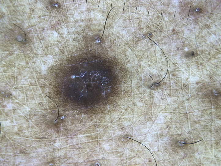
Dermatofibroma on right thigh of 64-year-old Black man, demonstrating lack of central white scar, fine reticulated pigment, and overlying scale. Photographed by Richard Usatine, MD.
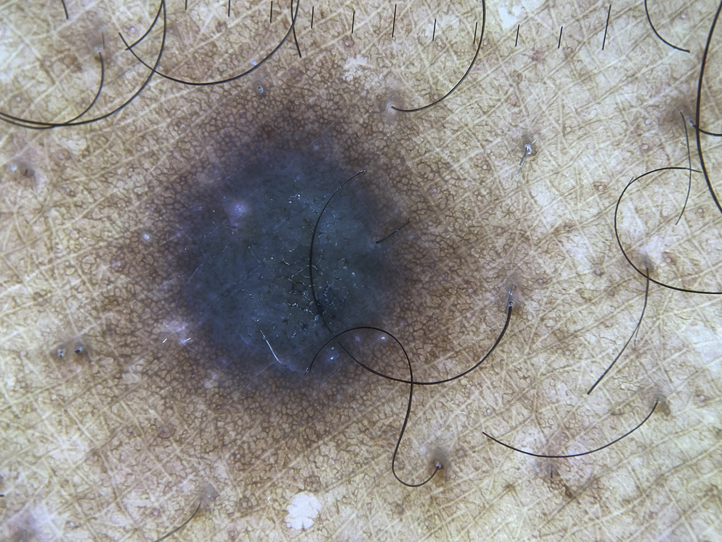
Dermatofibroma on left thigh of 64-year-old Black man, demonstrating blue-gray color, peripheral reticulation, perifollicular hypopigmentation, and lack of central scar. Photographed by Richard Usatine, MD.
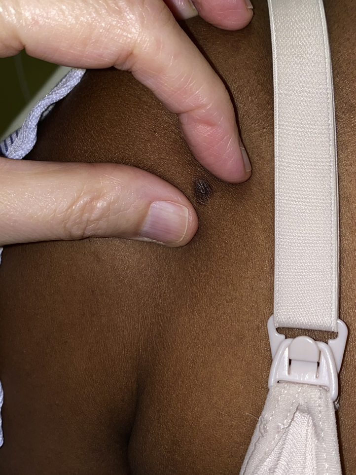
Dermatofibroma on chest of 56-year-old Black man, demonstrating lack of central scar, uniformly distributed brown network-like appearance, fine reticulation around the border, and a pink blush tone. Photographed by Elizabeth Seiverling, MD.
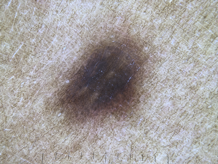
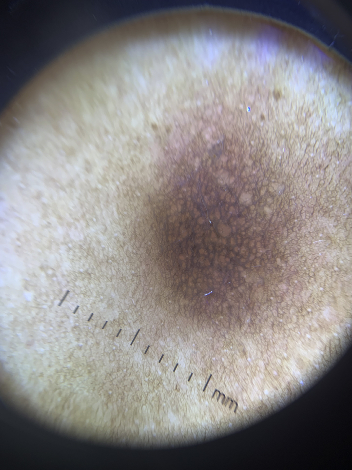
Dermatofibroma found on shoulder of 37-year-old Black woman, showing fine peripheral network, 6 patchy areas representing perifollicular hypopigmentation, and pink blush. Photographed by Richard Usatine, MD.
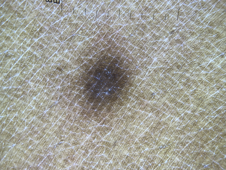
Dermatofibroma on leg in 37-year-old Black woman, demonstrating lack of central scar, delicate network lines in periphery, and perifollicular hypopigmentation. Photographed by Richard Usatine, MD.
Similar articles
-
Atrophic Dermatofibroma: A Unique Dermatofibroma Variant.Cureus. 2021 Apr 19;13(4):e14570. doi: 10.7759/cureus.14570. Cureus. 2021. PMID: 34035995 Free PMC article.
-
Clinical and histological patterns of dermatofibroma without gross skin surface change: A comparative study with conventional dermatofibroma.Indian J Dermatol Venereol Leprol. 2015 May-Jun;81(3):263-9. doi: 10.4103/0378-6323.154795. Indian J Dermatol Venereol Leprol. 2015. PMID: 25851763
-
Dermoscopy of dermatofibromas: a prospective morphological study of 412 cases.Arch Dermatol. 2008 Jan;144(1):75-83. doi: 10.1001/archdermatol.2007.8. Arch Dermatol. 2008. PMID: 18209171
-
Unique histopathologic features of the eyelid dermatofibroma.Orbit. 2019 Aug;38(4):274-278. doi: 10.1080/01676830.2018.1513045. Epub 2018 Sep 5. Orbit. 2019. PMID: 30183445 Review.
-
Multiple, clustered dermatofibroma: a rare clinical variant of dermatofibroma.J Cutan Med Surg. 2003 Nov-Dec;7(6):455-7. doi: 10.1007/s10227-002-0155-9. J Cutan Med Surg. 2003. PMID: 14605916 Review.
Cited by
-
Clinical and Dermoscopic Patterns of Basal Cell Carcinoma and Its Mimickers in Skin of Color: A Practical Summary.Medicina (Kaunas). 2024 Aug 24;60(9):1386. doi: 10.3390/medicina60091386. Medicina (Kaunas). 2024. PMID: 39336428 Free PMC article. Review.
-
Dermoscopy of Cutaneous Neoplasms in Skin of Color - A Systematic review by the International Dermoscopy Society "Imaging in Skin of Color" Task Force.Dermatol Pract Concept. 2023 Oct 1;13(4 S1):e2023308S. doi: 10.5826/dpc.1304S1a308S. Dermatol Pract Concept. 2023. PMID: 37874990 Free PMC article. Review.
References
Publication types
Grants and funding
LinkOut - more resources
Full Text Sources
Miscellaneous