. 2022 Jan 25;25(2):103812.
doi: 10.1016/j.isci.2022.103812. eCollection 2022 Feb 18.

SARM1 is a multi-functional NAD(P)ase with prominent base exchange activity, all regulated bymultiple physiologically relevant NAD metabolites

Affiliations

SARM1 is a multi-functional NAD(P)ase with prominent base exchange activity, all regulated bymultiple physiologically relevant NAD metabolites

Carlo Angeletti et al. iScience. .

Abstract

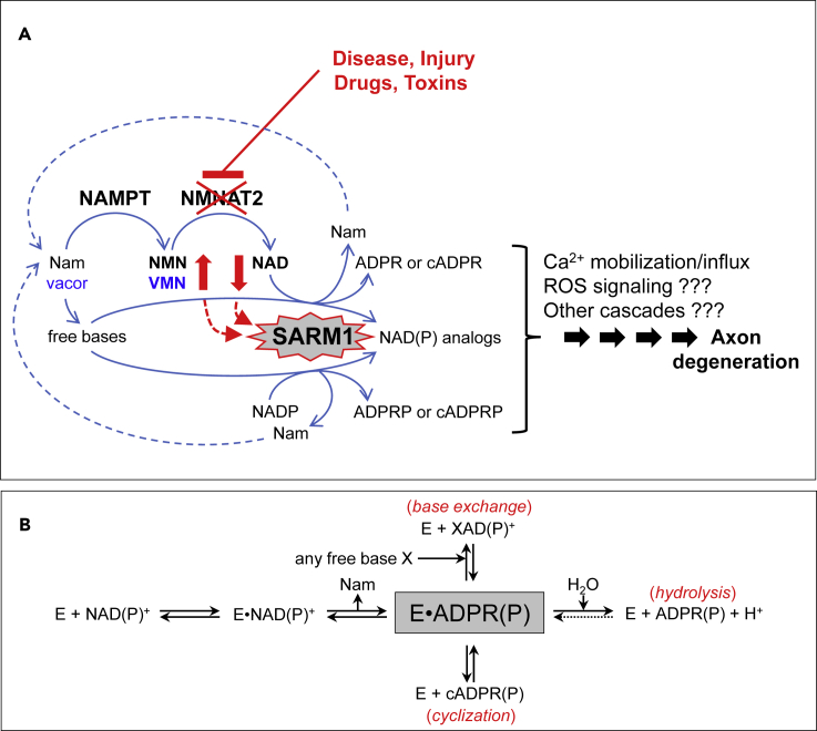
SARM1 is an NAD(P) glycohydrolase and TLR adapter with an essential, prodegenerative role in programmed axon death (Wallerian degeneration). Like other NAD(P)ases, it catalyzes multiple reactions that need to be fully investigated. Here, we compare these multiple activities for recombinant human SARM1, human CD38, and Aplysia californica ADP ribosyl cyclase. SARM1 has the highest transglycosidation (base exchange) activity at neutral pH and with some bases this dominates NAD(P) hydrolysis and cyclization. All SARM1 activities, including base exchange at neutral pH, are activated by an increased NMN:NAD ratio, at physiological levels of both metabolites. SARM1 base exchange occurs also in DRG neurons and is thus a very likely physiological source of calcium-mobilizing agent NaADP. Finally, we identify regulation by free pyridines, NADP, and nicotinic acid riboside (NaR) on SARM1, all of therapeutic interest. Understanding which specific SARM1 function(s) is responsible for axon degeneration is essential for its targeting in disease.

Keywords: Biological sciences; Molecular physiology; Neuroscience.


Conflict of interest statement

Funding for academic research from AstraZeneca and M.P.C. is a consultant for Nura Bio.

Figures

None
Graphical abstract
Figure 1
Figure 1
SARM1 is a central executioner of axon death via a multicomposite catabolic reaction typical of multifunctional NAD(P) glycohydrolases (A) Programmed axon degeneration is a widespread axon death mechanism driven by activation of SARM1 NAD(P)ase and prevented by its negative regulator NMNAT2, a short half-life enzyme essential to convert NMN into NAD. NMNAT2 forms together with the upstream enzyme NAMPT a key two-step pathway for NAD salvage in mammals that also provides metabolic conversion of the prodrug vacor into the neurotoxic, recently discovered, VMN intermediate. When NMNAT2 in axons becomes depleted or inactive, both NMN and VMN rise and NAD declines concomitantly; these fluctuations trigger SARM1 NAD(P)ase and, as shown here also base exchange, and thus initiate a signaling cascade that culminates into axon death. The early activation mechanism and key components of this process are highlighted in red and in blue for vacor toxicity. (B) Ordered Uni-Bi reaction mechanism of multifunctional NAD(P)ases (EC 3.2.2.6) like SARM1. From the substrate NAD(P), the pyridine moiety, Nam, is released first and then two distinct ADP ribosylated products arising from a single common intermediate (gray boxed). These may be ADPR(P) via classical hydrolysis or cADPR(P) via anhydrous cyclization. A third product XAD(P) represents any dinucleotides formed instead via transglycosidation (EC 2.4.99.20), a reaction of base exchange that replaces the pyridine moiety of the substrate with any related free base available.
Figure 2
Figure 2
Preliminary characterization of selected multifunctional NAD(P)ases (A) C18-HPLC UV profiles from time-course analyses of the three indicated multifunctional NAD(P)ase enzymes studied, all assayed with 250 μM NAD at 25°C under similar rates of substrate consumption. Both ADPR and cADPR products accumulating into diverse proportions are highlighted while unmarked in between, the substrate NAD peak declines in parallel and proportionally by time. (B) pH studies carried out by HPLC assays in universal buffer (Tris/Bis-Tris/Na-acetate) for 1 h at 25°C using 0.25 mM NAD and 7 μg/mL hSARM1 or 0.5 mM NAD and 0.07 μg/mL CD38 or 6 mM NAD and 0.08 μg/mL Aplysia cyclase. Data are Mean ± SEM from n = 3 and are normalized to relative maxima of each curve (see asterisks). (C) Optimum temperatures. HPLC assays were set for 1 h using 0.25 mM NAD and 11 μg/mL hSARM1 or 0.5 mM NAD and 0.1 μg/mL CD38 or 2.5 mM NAD and 0.14 μg/mL Aplysia cyclase. Data are Mean ± SEM from n = 3 and are normalized as in (B). (D and E) Preferred substrates (D) and effects of metal ions (E). Various dinucleotides (250 μM each) or metal ions (1 mM each) were assayed at 25°C by HPLC for 2–6 h using 5.5 μg/mL hSARM1 or 0.1 μg/mL CD38 or 0.028 μg/mL Aplysia cyclase. Data are Mean ± SEM from n ≥ 2. Dinucleotide analogs indicated are: NGD, nicotinamide guanine dinucleotide; NHD, nicotinamide hypoxanthine dinucleotide; AcPyAD, 3-acetylpyridine adenine dinuclotide; NaAD, nicotinic acid adenine dinuclotide; NaADP, NaAD phosphate; αNAD, alpha-NAD; ϵNAD, nicotinamide 1,N6-etheno adenine dinucleotide; VAD, vacor adenine dinucleotide; NADH or NADPH, reduced NAD or NADP. See also Figures S1–S3.
Figure 3
Figure 3
NAD and NADP kinetics by SARM1 in comparison to other multifunctional NAD(P)ases and under allosteric triggering by NMN (A) NAD kinetics of the three multifunctional NAD(P)ases. Initial rates were measured by HPLC using 3 μg/mL of hSARM1 or 0.05 μg/mL of CD38 or 0.02 μg/mL of Aplysia cyclase. Assays were carried out at 25°C for 30 min. Data are Mean ± SEM from n ≥ 3. Insets, double reciprocal plot analyses. Dotted lines, best fitting analyses carried out using the Equations 1 and 3 in STAR methods. The recalculated kinetic parameters are shown in Table 1. (B) NAD kinetics of the human SARM1 fragment SAM-TIR with constitutive NAD(P)ase that is not inducible by NMN because of the lack of the auto-inhibitory N-terminal regulatory domain ARM. HPLC assays were carried out at 25°C for 1 h using 2.75 μg/mL of enzyme per mix. Data are Mean ± SEM from n = 4. Inset, double reciprocal plot analysis. Dotted line, best fitting results from Equation 2 in STAR methods (see also recalculated parameters in Table 1). (C) NAD kinetics of hSARM1 full length (3 μg/mL per mix as in (A)) at increasing micromolar concentrations of the allosteric regulator NMN. Best fitting analysis was carried out on individual curves using Equation 3 in STAR methods to calculate the kinetic parameters shown in Table 1, and the relative curve maxima that were subsequently re-plotted on the flanking graph. Their linear relationship with the trigger amount in each curve indicates competition between NMN and NAD for opposing regulation of hSARM1 NADase. (D) NADP kinetics presented as for NAD in A above but done by assaying 14.6 μg/mL of hSARM1 or 0.05 μg/mL of CD38 or 0.07 μg/mL of Aplysia cyclase at 25°C for 60–120 min. Data are Mean ± SEM from n = 3. (E) NADP kinetics as in B above but done by assaying 1.9 μg/mL of SAM-TIR at 25°C for 1 h. Data are Mean ± SEM from n = 2. (F) NADP kinetics as in C above (3.7 μg/mL of hSARM1 full length per mix) at increasing micromolar concentrations of NMN. Flanking graph, best fitting maxima re-plotted showing NADP effects unrelated to allosteric triggering by NMN of hSARM1 NADPase. See also Figures S1 and S2.
Figure 4
Figure 4
Base exchange reactions typically catalyzed by SARM1 and other multifunctional NAD(P)ases at neutral pH (A and B) Rates were measured in 50 mM HEPES/NaOH pH 7.5 at 25°C by HPLC using 0.05 μg/mL of CD38 or 0.1 μg/mL of Aplysia cyclase or 0.5 μg/mL of SARM1 SAM-TIR (upper panel A) or 7 μg/mL of SARM1 full length (bottom panel B). Both NAD and NADP substrates were fixed at 250 μM. The free bases 3-acetylpyridine (AcPyr) and nicotinic acid (Na) were added at 2 mM final. Vacor was added at 0.5 mM given its low solubility at physiological pH. The assay in B (left panel) is also shown in Supplementary (see Figure S4) and was replicated in the presence of two known allosteric regulators of SARM1, NMN 0.2 mM and VMN 0.05 mM (B, middle and right panels), leading both to a maximum triggering effect on SARM1 activity at this concentration as reported (Loreto et al., 2021). Multiple time stops from individual assays as above were analyzed for linearity, then extents of hydrolysis (white bars), cyclization (red bars), or base exchange (black bars) were calculated from each corresponding product as shown in Figure 1B and STAR methods. In detail, base exchanges led to form AcPyrAD from AcPyr, VAD from vacor, NaAD from Na in the presence of NAD or AcPyrADP from AcPyr, VADP from vacor, NaADP from Na in the presence of NADP (see Figure S4). Rates (Mean ± SEM, n = 2) are shown in histograms for comparison, referred to either NAD or NADP alone controls (arbitrarily fixed to 1). The whole dataset is also shown in Table S1. Asterisks (∗) indicate conditions where base exchange was below detection. See also Figures S3–S5, and Table S1
Figure 5
Figure 5
First evidence for SARM1-dependent base exchanges in DRG neurons (A) Quantification of the degeneration index and representative images of neurites from wild-type and Sarm1−/− DRG explant cultures treated with 3-acetylpyridine (AcPyr) at the timepoints indicated (mean ± SD; n = 3). (B) Typical C18-HPLC UV profiles of the nucleotide extract obtained from wild-type and Sarm1−/− DRG explant cultures following treatment for 4 h with AcPyr 250 μM or vehicle. The compounds of interest are indicated. (C) Measured levels of the different nucleotides extracted and analyzed as above (mean ± SD; n = 3). Data are normalized to the protein amount extracted in parallel. Asterisks (∗) indicate levels below detection.
Figure 6
Figure 6
Selective inhibition by pyridine ribosides on multifunctional NAD(P)ase members (A) NADase inhibition at 200 μM fixed NR, NaR, or VR of the indicated enzyme species. Assays were carried out by HPLC at 25°C for 1 h using 0.25 mM of NAD and 13 μg/mL of SARM1 or 0.25 mM of NAD and 3.9 μg/mL of SAM-TIR or 0.25 mM of NAD and 0.1 μg/mL of CD38 or 1 mM of NAD and 0.02 μg/mL of Aplysia cyclase. Data are Mean ± SEM from n ≥ 4, normalized to NAD alone controls. (B) NADase inhibition at variable NaR or VR of SARM1 full length and SAM-TIR (assayed as in A above). Data are Mean ± SEM from n = 3. Dotted lines, best fitting analyses carried out using Equation 4 in STAR methods with the calculated IC50 values highlighted. (C and D) NADase inhibition kinetics under variable substrate NAD and at various fixed concentrations of either NaR or VR as indicated. SARM1 SAM-TIR (0.6 μg/mL in mix) was assayed by HPLC for 1–3 h at 25°C. The graph shows also Lineweaver-Burk plots (C and D middle) and slope replots (C and D right) indicating, respectively, inhibition type and Ki for both NaR and VR and the calculated number (n) of inhibitor molecules that bind to the enzyme. Best fitting analysis was done with Equation 5 in STAR methods.

References

    1. Aksoy P., White T.A., Thompson M., Chini E.N. Regulation of intracellular levels of NAD: a novel role for CD38. Biochem. Biophys. Res. Commun. 2006;345:1386–1392. doi: 10.1016/j.bbrc.2006.05.042. - DOI - PubMed
    1. Anderson G.D., Peterson T.C., Farin F.M., Bammler T.K., Beyer R.P., Kantor E.D., Hoane M.R. The effect of nicotinamide on gene expression in a traumatic brain injury model. Front. Neurosci. 2013;7:21. doi: 10.3389/fnins.2013.00021. - DOI - PMC - PubMed
    1. Belenky P., Christensen K.C., Gazzaniga F., Pletnev A.A., Brenner C. Nicotinamide riboside and nicotinic acid riboside salvage in fungi and mammals. Quantitative basis for Urh1 and purine nucleoside phosphorylase function in NAD+ metabolism. J. Biol. Chem. 2009;284:158–164. doi: 10.1074/jbc.M807976200. - DOI - PMC - PubMed
    1. Berger F., Ramirez-Hernandez M.H., Ziegler M. The new life of a centenarian: signalling functions of NAD(P) Trends Biochem. Sci. 2004;29:111–118. doi: 10.1016/j.tibs.2004.01.007. - DOI - PubMed
    1. Berthelier V., Tixier J.M., Muller-Steffner H., Schuber F., Deterre P. Human CD38 is an authentic NAD(P)+ glycohydrolase. Biochem. J. 1998;330:1383–1390. doi: 10.1042/bj3301383. - DOI - PMC - PubMed