. 2022 Feb 2;25(2):103860.
doi: 10.1016/j.isci.2022.103860. eCollection 2022 Feb 18.

Zrsr2 and functional U12-dependent spliceosome are necessary for follicular development

Affiliations

Zrsr2 and functional U12-dependent spliceosome are necessary for follicular development

Isabel Gómez-Redondo et al. iScience. .

Abstract

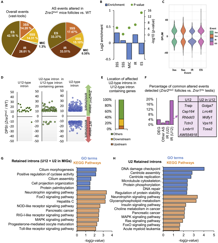
ZRSR2 is a splicing factor involved in recognition of 3'-intron splice sites that is frequently mutated in myeloid malignancies and several tumors; however, the role of mutations of Zrsr2 in other tissues has not been analyzed. To explore the biological role of ZRSR2, we generated three Zrsr2 mutant mouse lines. All Zrsr2 mutant lines exhibited blood cell anomalies, and in two lines, oogenesis was blocked at the secondary follicle stage. RNA-seq of Zrsr2 mu secondary follicles showed aberrations in gene expression and showed altered alternative splicing (AS) events involving enrichment of U12-type intron retention (IR), supporting the functional Zrsr2 action in minor spliceosomes. IR events were preferentially associated with centriole replication, protein phosphorylation, and DNA damage checkpoint. Notably, we found alterations in AS events of 50 meiotic genes. These results indicate that ZRSR2 mutations alter splicing mainly in U12-type introns, which may affect peripheral blood cells, and impede oogenesis and female fertility.

Keywords: Biological sciences; Molecular biology; Transcriptomics.


Conflict of interest statement

The authors declare no competing interests.

Figures

None
Graphical abstract
Figure 1
Figure 1
Diagrams of Zrsr2 gene structure and mutations generated in this study and their expression (A) Diagram of the Zrsr2 gene with its functional domains: ZF1 and ZF2 (zinc finger domains 1 and 2), RRM (RNA recognition motif), and RS (arginine-serine rich). The red lightning indicates the target region for each different mutation. The protein sizes and structures produced by the different mutations (Zrsr2muA, Zrsr2muB, and Zrsr2muC) are represented in the lower panels, shaded in pink. The position of the primers used for Figure 1C (2-3F and 7R) and Figure 1C (9F and 11R) are indicated. (B) Western blot analysis of ZRSR2 protein expression in spleen of WT and the three mutant mice, using ACTB as a loading control. The positions of molecular mass markers are indicated on the left. Blue arrows indicate ZRSR2 mutant protein produced by different alternative splicing events. (C) Expression level of Zrsr2 mRNA in ovaries from 3-month-old females of the three mutant lines and WT mice determined by RT-qPCR. Biological triplicate data for qPCR are presented as mean ± SEM. Bars with different superscripts differ significantly (p < 0.05). (D) Non-quantitative RT-PCR analysis of Zrsr2 in WT and Zrsr2muA, Zrsr2muB, and Zrsr2muC mice. The right panel shows a diagram of the affected exons in Zrsr2muC mice. Isoforms were confirmed by sequencing.
Figure 2
Figure 2
Ovarian morphology and histology and follicle count in WT and Zrsr2 mutant mice (A) WT and Zrsr2 mutant ovaries from the three lines at 5-, 8-, and 11-week-old mice. Scale bar, 200 μm. (B) Hematoxylin and eosin-stained sections of WT, Zrsr2muA, Zrsr2muB, and Zrsr2muC ovaries from 5- and 8-week-old mice. Scale bar, 200 μm. (C) Bar graph representing the ovary length of WT, Zrsr2muA, Zrsr2muB, and Zrsr2muC ovaries from 5-, 8- and 11-week-old mice (n = 5 per line). Biological triplicate data for qPCR are presented as mean ± SEM. Bars with different superscripts differ significantly (p < 0.05). (two-way ANOVA followed by Tukey’s post hoc test). (D) Primary, secondary, and antral follicles count in dissected ovaries from 8- to 10-week-old females from the WT and the three Zrsr2mu lines (n = 5 per line) injected with pregnant mare’s serum gonadotropin (PMSG). Biological triplicate data for qPCR are presented as mean ± SEM. Bars with different superscripts differ significantly (p < 0.05) (two-way ANOVA followed by Tukey’s post hoc test).
Figure 3
Figure 3
Peripheral blood cell counts Boxplot showing the most representative parameters of each cell type series (white blood cells, red blood cells, and platelets). WBC, white blood cells; LYM, lymphocytes; MON, monocytes; GRAN, granulocytes; RBC, red blood cells; MCV, erythrocyte volume; MCH, hemoglobin content; RDW, variation coefficient; PLT, platelets; MPV, mean platelet volume; PDW, heterogeneity; PCT, percentage of blood occupied by platelets. Bars with different superscripts differ significantly (p < 0.05) (one-way ANOVA followed by Tukey’s post hoc test).
Figure 4
Figure 4
Zrsr2 expression profile and RNA-seq analysis of WT and Zrsr2muC follicles (A) Relative expression levels of Zrsr2 in developing follicles; biological triplicate results presented as the mean ± SEM. Bars with different superscripts differ significantly (p < 0.05 by two-way ANOVA followed by Tukey’s post hoc test). (B) Pie charts show the proportion of up-regulated and down-regulated genes as well as the cellular components of the DEGs. (C) Volcano plot of differentially expressed genes between Zrsr2muC and wild-type samples, in which genes with an false discovery rate value under 0.01 and a fold change over 2 are represented in red. The genes with the greatest fold change and/or significant difference are indicated. Four genes of calcium-binding protein are highlighted surrounded by blue. (D) Bar plot of the number of genes belonging to the three main gene types in the set of DEGs. (E) Number of DEGs that are expressed preferentially in the oocyte, in the granulosa cells or both (oocyte expression data: GEO-GSE111687, granulosa cells expression data: GEO-GSE158218). Missing genes in those datasets are indicated as “Non-classified.” (F) Significantly enriched molecular functions terms in down-regulated transcripts in the Zrsr2muC follicles. (G) Significantly enriched biological process terms in down-regulated transcripts in the Zrsr2muC follicles. (H) RNA-seq coverage plot of Zrsr2 gene (top panel) representing the WT data (green) and the Zrsr2muC data (red). The lower panel shows a zoom of the last exons of Zrsr2, showing the coverage and the splice junctions (shown in blue) of WT and Zrsr2muC data.
Figure 5
Figure 5
RNA-seq analysis of alternative splicing in Zrsr2muC follicles (A) Distribution of categories of alternative splicing (AS) events differing in Zrsr2muC secondary follicles versus WT. Percentages of each class of event in vast-tools (VastDB annotation) and in Zrsr2muC follicles are indicated. 3SS, alternative 3′ splice sites; A5SS, alternative 5′ splice sites; ES, exon skipping; MIC, alternative micro cassette exon ≤15 nucleotides; IR, intron retention. (B) Enrichment of differentially spliced events in Zrsr2muC secondary follicles versus WT with respect to the total number of events annotated in the mouse genome. MIC values are chopped for clarity purposes. (C) Violin plots of AS events differing in Zrsr2muC secondary follicles versus WT. (D) Differences in intron retention events detected in Zrsr2mu compared with WT follicles (measured as delta percent-spliced-in [DPSI] ratio of intron read counts in Zrsr2muC versus WT for different intron categories, as indicated). (E) Distributions of the locations of affected U2-type introns in Zrsr2muC follicles relative to U12-type introns present in the same gene. (F) Percentage of events altered in Zrsr2muC compared with WT follicles that were also detected in Zrsr1mu testis (Horiuchi et al., 2018). DEG, differentially expressed genes; Other AS: 3SS, 5SS, ES, and MIC; IR (U2), U2-type introns that showed IR; IR (U12), U12- and U2-type introns located in genes with U12 that showed IR. The table on the right shows the genes with common IR (U12) events between the two experiments. (G) Significantly enriched GO terms and KEGG pathways in MIGs showing retention in U12- or U2-type introns in genes containing U12-type introns in Zrsr2muC. (H) Significantly enriched KEGG pathways in MIGs showing retention in up-regulated U2 introns in Zrsr2muC

Comment in

  • Differential alternative splicing analysis links variation in ZRSR2 to a novel type of oral-facial-digital syndrome.
    Hannes L, Atzori M, Goldenberg A, Argente J, Attie-Bitach T, Amiel J, Attanasio C, Braslavsky DG, Bruel AL, Castanet M, Dubourg C, Jacobs A, Lyonnet S, Martinez-Mayer J, Pérez Millán MI, Pezzella N, Pelgrims E, Aerden M, Bauters M, Rochtus A, Scaglia P, Swillen A, Sifrim A, Tammaro R, Mau-Them FT, Odent S, Thauvin-Robinet C, Franco B, Breckpot J. Hannes L, et al. Genet Med. 2024 Apr;26(4):101059. doi: 10.1016/j.gim.2023.101059. Epub 2023 Dec 27. Genet Med. 2024. PMID: 38158857

References

    1. Abou Alezz M., Celli L., Belotti G., Lisa A., Bione S. GC-AG introns features in long non-coding and protein-coding genes suggest their role in gene expression regulation. Front Genet. 2020;11:488. - PMC - PubMed
    1. Aken B.L., Ayling S., Barrell D., Clarke L., Curwen V., Fairley S., Fernandez Banet J., Billis K., Garcia Giron C., Hourlier T., et al. The Ensembl Gene Annotation System. Database (Oxford) 2016;2016:baw093. - PMC - PubMed
    1. Alen F., Gomez-Redondo I., Rivera P., Suarez J., Ramos-Ibeas P., Pericuesta E., Fernandez-Gonzalez R., Perez-Cerezales S., Horiuchi K., Orio L., et al. Sex-dimorphic behavioral alterations and altered neurogenesis in U12 intron splicing-defective Zrsr1 mutant mice. Int. J. Mol. Sci. 2019;20:3543. - PMC - PubMed
    1. Alioto T.S. U12DB: a database of orthologous U12-type spliceosomal introns. Nucleic Acids Res. 2007;35:D110–D115. - PMC - PubMed
    1. Anders S., Pyl P.T., Huber W. HTSeq--a Python framework to work with high-throughput sequencing data. Bioinformatics. 2015;31:166–169. - PMC - PubMed