. 2022 Feb 8:13:100213.
doi: 10.1016/j.mtbio.2022.100213. eCollection 2022 Jan.

Vapor construction and modification of stem cell-laden multicomponent scaffolds for regenerative therapeutics

Affiliations

Vapor construction and modification of stem cell-laden multicomponent scaffolds for regenerative therapeutics

Yu-Chih Chiang et al. Mater Today Bio. .

Abstract

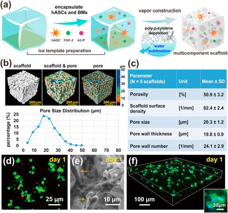
Tissue engineering based on the combined use of isolated cells, scaffolds, and growth factors is widely used; however, the manufacture of cell-preloaded scaffolds faces challenges. Herein, we fabricated a multicomponent scaffold with multiple component accommodations, including bioactive molecules (BMs), such as fibroblast growth factor-2 (FGF-2) and l-ascorbic acid 2-phosphate (A2-P), and living cells of human adipose-derived stem cells (hASCs), within one scaffold construct. We report an innovative fabrication process based on vapor-phased construction using iced templates for vapor sublimation. Simultaneously, the vaporized water molecules were replaced by vapor deposition of poly-p-xylylene (PPX, USP Class VI, highly compatible polymer, FDA-approved records), forming a three-dimensional and porous scaffold matrix. More importantly, a multicomponent modification was achieved based on using nonvolatile solutes, including bioactive molecules of FGF-2 and A2-P, and living cells of hASCs, to prepare iced templates for sublimation. Additionally, the fabrication and construction resulted in a multicomponent scaffold product comprising the devised molecules, cells, and vapor-polymerized poly-p-xylylene as the scaffold matrix. The clean and dry fabrication process did not require catalysts, initiators or plasticizers, and potentially harmful solvents, and the scaffold products were produced in simple steps within hours of the processing time. Cell viability analysis showed a high survival rate (approximately 86.4%) for the accommodated hASCs in the fabricated scaffold product, and a surprising multilineage differentiation potential of hASCs was highly upregulated because of synergistic guidance by the same accommodated FGF-2 and A2-P components. Proliferation and self-renewal activities were also demonstrated with enhancement of the multicomponent scaffold product. Finally, in vivo calvarial defect studies further revealed that the constructed scaffolds provided blood vessels to grow into the bone defect areas with enhancement, and the induced conduction of osteoblast growth also promoted bone healing toward osseointegration. The reported scaffold construction technology represents a prospective tissue engineering scaffold product to enable accommodable and customizable versatility to control the distribution and composition of loading delicate BMs and living hASCs in one scaffold construct and demonstrates unlimited applications in tissue engineering repair and regenerative medicine applications.

Keywords: Growth factor; Human adipose tissue-derived stem cell; Multifunctional scaffold; Regenerative medicine; Vapor construction.


Conflict of interest statement

The authors declare that they have no known competing financial interests or personal relationships that could have appeared to influence the work reported in this paper.

Figures

Image 1
Graphical abstract
Fig. 1
Fig. 1
Scaffold fabrication and characterization. (a) Proposed manufacturing process using controlled mass transport during vapor-phased sublimation/deposition to construct a multicomponent scaffold and simultaneously accommodated human adipose-derived stem cells (hASCs) and multiple bioactive molecules of fibroblast growth factor-2 (FGF-2) and l-ascorbic acid 2-phosphate (A2-P). (b) Micro-CT imaging of the manufactured scaffold showed a porous structure and interconnected pores. The measured pore size distribution ranged from 5 to 50 ​μm. (c) Scaffold morphometrical properties (N ​= ​5 scaffolds) assessed by micro-CT analysis. (d) The cell viability assessed by LIVE/DEAD staining analysis at day 1 showed that approximately 86.4% of the accommodated hASCs were alive and emitted green fluorescence in the fabricated scaffold. Dead cells were stained in red for comparison. (e) The SEM image of the interior structure showed that the accommodated hASCs adhered and grew on the manufactured porous scaffold. (f) The 3D confocal micrograph further showed that the accommodated hASCs were evenly distributed in the fabricated scaffold. hASCs were fluorescence labeled with Alexa Fluor® 488-conjugated phalloidin to stain the cytoskeleton (green) and DAPI to stain the nucleus (blue); a high resolution and magnified image of stained cells is shown in the inset.
Fig. 2
Fig. 2
Cell proliferation behavior analysis. (a) Cell morphology and growth pattern on the fabricated scaffolds that accommodate specific bioactive molecules (BMs) and living hASCs. The viable and proliferating hASCs (green channel) were visualized by LIVE/DEAD staining at days 1, 5, and 10. The red background was mainly the reflection of the noise signal on the surface of the scaffold. Compared with other studied scaffolds, the scaffold containing FGF-2 and A2-P (i.e., hASC-BM scaffold) had the best effect on enhancing cell proliferation. (b) The metabolic activity curves of the accommodated hASCs were determined using the MTT assay. (c) The doubling time required for the logarithmic growth of the accommodated cell population was further calculated and compared. The statistical data are expressed as mean values ​± ​standard deviation based on three independent samples (∗: p-value < 0.05; ∗∗: p-value < 0.01; ∗∗∗: p-value < 0.001, compared to the hASC scaffold group).
Fig. 3
Fig. 3
Cell self-renewal marker expression. (a) Immunofluorescence of stem cell pluripotent markers Oct-4, Sox-2 and Nanog showing the stem cell guidance property on the fabricated scaffolds that accommodate specific bioactive molecules (BMs) and living hASCs. After 7 days of culture, the fabricated scaffold containing A2-P and FGF-2 molecules (i.e., hASC-BM scaffold) enhanced the self-renewal activities of accommodated hASCs. These hASCs were harvested from the scaffolds by trypsinization and analyzed with approximately the same number of cells. All the nuclei were counterstained with DAPI (blue channel) to guide imaging. For comparison, a control scaffold (i.e., hASC scaffold) containing hASCs within the pure PPX matrix was used in parallel. (b) Western blot analysis of Oct4, Sox2, and Nanog proteins. β-Actin served as an internal protein loading control. (c) Quantification of the band intensity using the densitometric analysis software ImageJ. The data are presented as the mean relative density ​± ​SD of three independent experiments; ∗: p-value < 0.05, compared to the hASC scaffold group.
Fig. 4
Fig. 4
Cell differentiation potential analysis. (a) The multilineage differentiation capabilities of hASCs, including osteogenesis, adipogenesis, and hepatogenesis, were confirmed by anti-osteocalcin immunofluorescence staining at day 21, Oil Red O staining at day 10, and anti-albumin immunofluorescence staining at day 21, respectively, on the fabricated scaffold accommodated with A2-P and FGF-2 molecules (i.e., hASC-BM scaffold) compared with a control scaffold (i.e., hASC scaffold) containing hASCs within the pure PPX matrix. The nucleus (blue) was counterstained with DAPI to indicate the cell position. (b) Quantification of fluorescence intensity using the image analysis software ImageJ. The data are presented as the mean relative fluorescence density ​± ​SD of three independent experiments; ∗: p-value < 0.05 and ∗∗: p-value < 0.01, compared with the control group.
Fig. 5
Fig. 5
Overview and micro-CT imaging evaluation of calvarial bone defects of animal study (rat). (a) Outline of the experimental design of rat calvarium. Bone trephine defects (5 ​mm in diameter critical-size) were made at the bilateral calvarium. (b) Calvarial bone defects were created, and the scaffold was implanted. (c) New bone formation evaluation of 3D micro-CT image reconstruction at 8 weeks postoperatively. PPX: poly-p-xylylene-based porous scaffold, PPX-hASC-BM: FGF-2/A2-P-treated and hASC accommodated PPX, Control: no scaffold used in trephine defects. (d) Bone volume and mineral density calculated from micro-CT 3D datasets using Amira software, asterisk (∗) indicated statistically significant difference (p ​< ​0.05). No significant difference was found in bone mineral density.
Fig. 6
Fig. 6
Representative micro-CT images and histology analyses of calvarial bone regeneration at trephine defects. (HE: hematoxylin and eosin stain; BSP: bone sialoprotein stain; F4/80: F4/80 antibody stain for macrophage markers). (a) No scaffold was used in the bone window at 8 weeks postsurgery. Micro-CT image showed the bone formed along the peripheral margin of the bone cavity wall. Mature laminar bone (LB) formed at the bottom of the bony cavity. Osteocytes (OCs) were clearly shown in woven bone. Fibrous connective tissue (CT) with hemorrhage was found at the margin of the bone cavity. Brain tissue (∗asterisk) at the bottom of the picture. BSP staining showed osteoblasts making bone in the bone marrow and along the woven bone. Few macrophages were detected (red arrows). (b) Nonfunctionalized pure PPX scaffold used in a bone window at 8 weeks postsurgery. The micro-CT image showed that new mineralized bone deposition occurred in the middle and peripheral areas of the trephine defect. Lamellar bone (LB) formed on the borders. Newly mineralized bone matrix (M) and bone marrow (BM) filled in the center. Red blood cells in capillaries (black arrow) formed in BM. Groups of osteoclasts were noted (white arrows). Promyelocytes and myelocytes were found in the bone marrow. Osteoclasts and osteoblasts surrounded a new bone matrix (NB). No inflammatory cell infiltration was observed (F4/80 detection) at this stage. (c) Proposed living cell- and bioactive molecule-loaded scaffolds (PPX-hASC-BM) used in the bone window at 8 weeks postsurgery. Micro-CT images showed new bone tissue formed in the middle of the trephine defect. More mature bone formed along the peripheral area of the bone cavity. A Haversian canal (black arrow) was formed in trabecular bone. Furthermore, numerous osteoprogenitor cells and collagen (∗asterisk) were observed between new bones in which osteocytes were lining. An intense reaction was observed between trabecular bones stained for BSP. A fragment of the PPX scaffold (red arrowheads) was surrounded by preosteoblasts (blue arrow) and osteoprogenitor cells (green arrow). No macrophage or lymphocyte/plasma cell infiltration was found (F4/80 detection).

References

    1. Arvidson K., et al. J. Cell Mol. Med. 2011;15(4):718. - PMC - PubMed
    1. Yamada Y., et al. Tissue Eng. 2004;10(5–6):955. - PubMed
    1. Whitaker M., et al. J. Pharm. Pharmacol. 2001;53(11):1427. - PubMed
    1. Wang Z., et al. NPG Asia Mater. 2017;9(10):e435. - PMC - PubMed
    1. De Witte T.M., et al. Regen Biomater. 2018;5(4):197. - PMC - PubMed

LinkOut - more resources