

Subcutaneous ICD Implantation and Catheter Ablation: A Step-Planned Approach for Ventricular Tachycardia Management in ARVC
- PMID: 35199014
- PMCID: PMC8855132
- DOI: 10.1016/j.jaccas.2021.12.007
Subcutaneous ICD Implantation and Catheter Ablation: A Step-Planned Approach for Ventricular Tachycardia Management in ARVC
Abstract
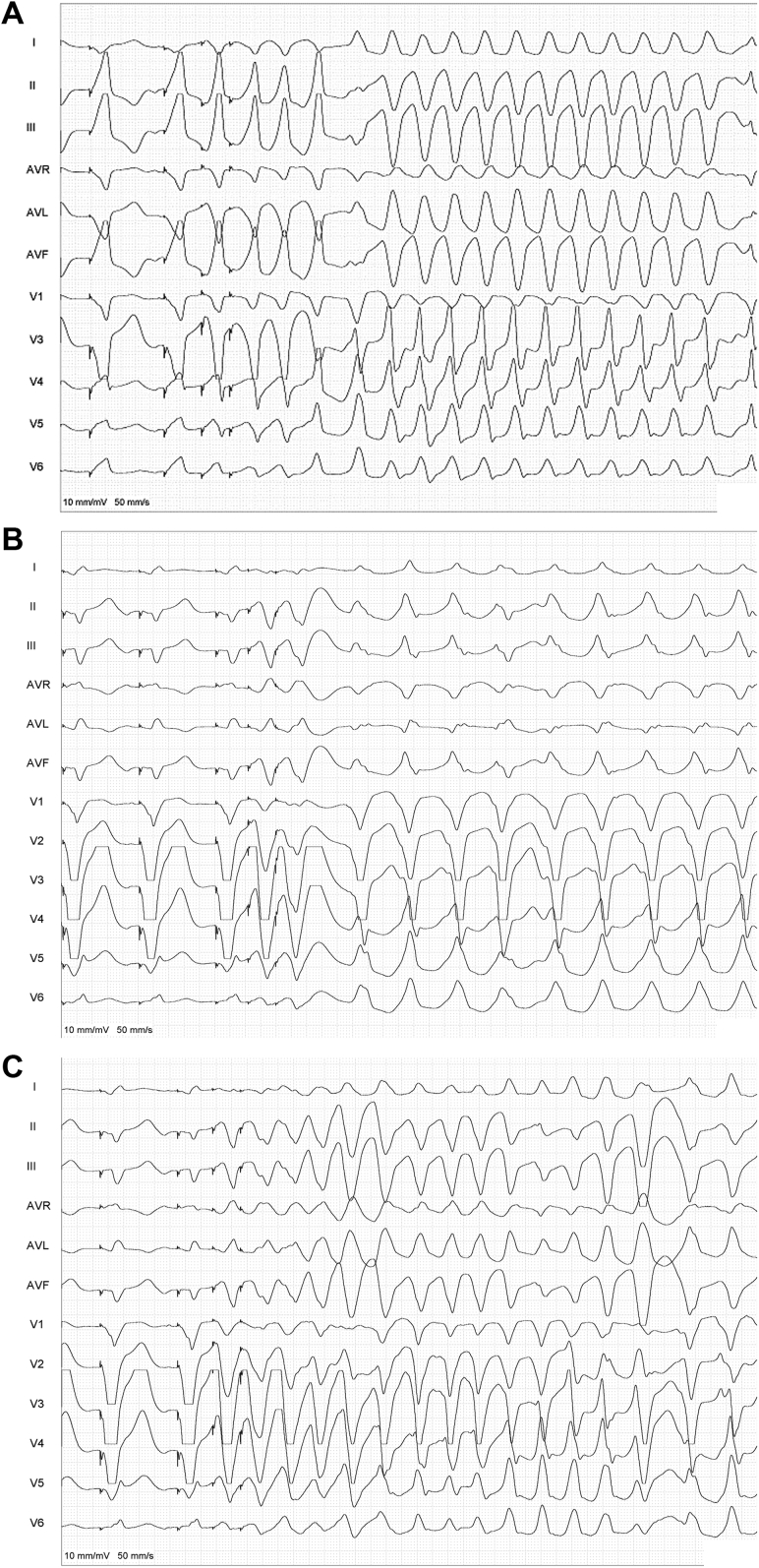
Secondary prevention of sudden cardiac death in the young patient with arrhythmogenic right ventricular cardiomyopathy and hemodynamically tolerated ventricular tachycardia is still a challenging field. We present a combined approach, including subcutaneous implantable cardioverter-defibrillator (ICD) and catheter ablation, as a promising treatment to prevent both ventricular tachycardia recurrences and ICD shocks. (Level of Difficulty: Intermediate.).
Keywords: 3D-EAM, 3-dimensional electroanatomical map; ARVC, arrhythmogenic right ventricular cardiomyopathy; ATP, antitachycardia pacing; CA, catheter ablation; CMR, cardiac magnetic resonance; ECG, electrocardiogram; ED, emergency department; EP, electrophysiological; ICD, implantable cardioverter-defibrillator; LBBB, left bundle branch block; LP, late potential; LV, left ventricle; NSVT, nonsustained ventricular tachycardia; PVS, programmed ventricular stimulation; RV, right ventricular; S-ICD, subcutaneous implantable cardioverter-defibrillator; VT, ventricular tachycardia; arrhythmogenic right ventricular cardiomyopathy; subcutaneous ICD; ventricular tachycardia ablation.
© 2022 The Authors.
Conflict of interest statement
The authors have reported that they have no relationships relevant to the contents of this paper to disclose.
Figures


References
-
- Caforio A.L.P., Pankuweit S., Arbustini E., et al. Current state of knowledge on aetiology, diagnosis, management, and therapy of myocarditis: a position statement of the European Society of Cardiology Working Group on Myocardial and Pericardial Diseases. Eur Heart J. 2013;34:2636–2648. - PubMed
-
- Towbin J.A., McKenna W.J., Abrams D.J., et al. 2019 HRS expert consensus statement on evaluation, risk stratification, and management of arrhythmogenic cardiomyopathy. Heart Rhythm. 2019;16(11):e301–e372. - PubMed
-
- Santangeli P., Zado E.S., Supple G.E., et al. Long-term outcome with catheter ablation of ventricular tachycardia in patients with arrhythmogenic right ventricular cardiomyopathy. Circ Arrhythm Electrophysiol. 2015;8:1413–1421. - PubMed
-
- Santangeli P., Hamilton-Craig C., Dello Russo A., et al. Imaging of scar in patients with ventricular arrhythmias of right ventricular origin: cardiac magnetic resonance versus electroanatomic mapping. J Cardiovasc Electrophysiol. 2011;22:1359–1366. - PubMed
Publication types
LinkOut - more resources
Full Text Sources
Research Materials
