. 2022 Feb 8:12:163-169.
doi: 10.1016/j.ibneur.2022.02.001. eCollection 2022 Jun.

Peripheral neuropathy in the pre-diabetic state of the type 2 diabetes mouse model (TSOD mice) involves TRPV1 expression in dorsal root ganglions

Affiliations

Peripheral neuropathy in the pre-diabetic state of the type 2 diabetes mouse model (TSOD mice) involves TRPV1 expression in dorsal root ganglions

Kyoko Shida et al. IBRO Neurosci Rep. .

Abstract

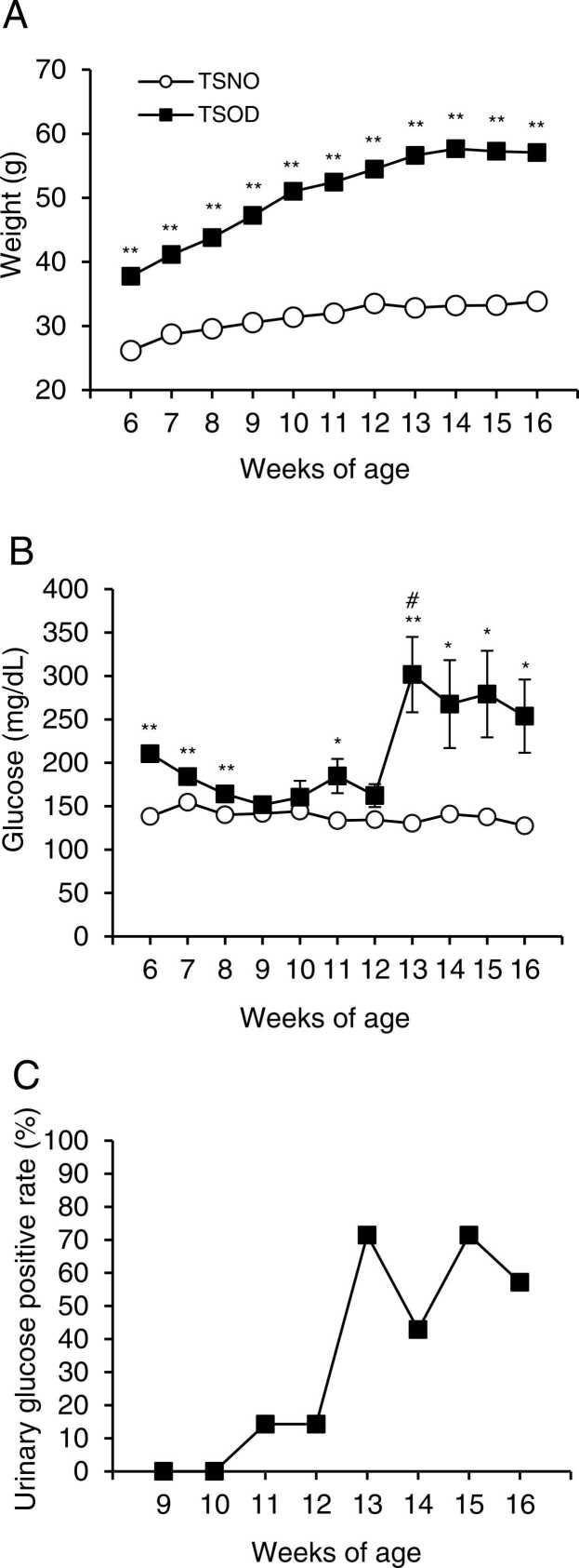
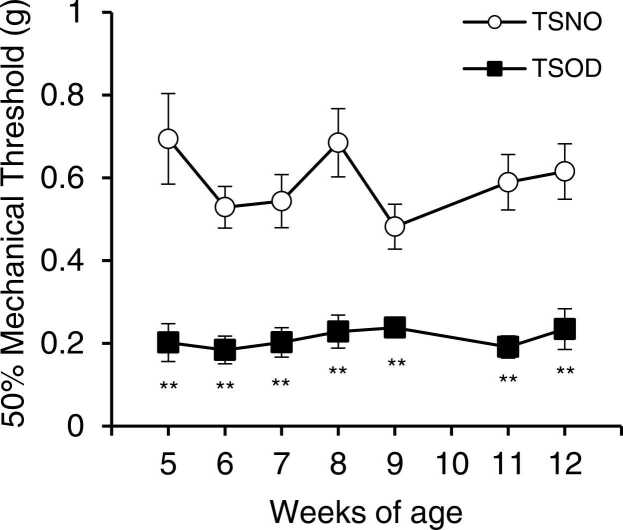
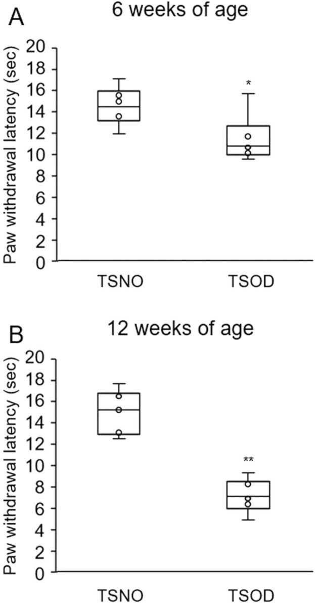
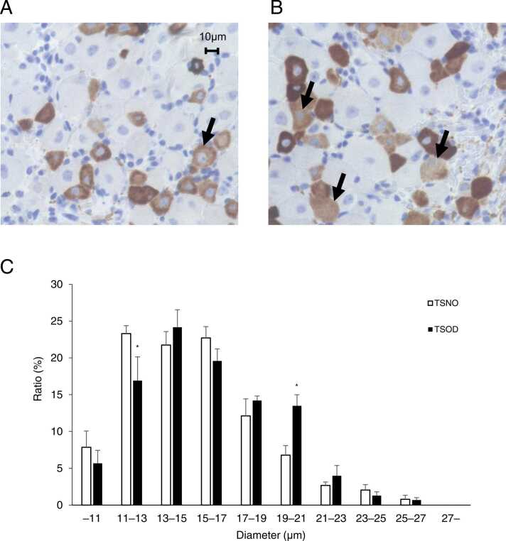
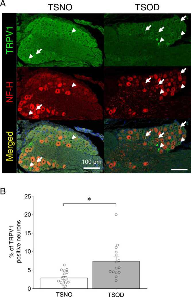
Peripheral neuropathy, which is a complication of diabetes mellitus (DM), is thought to occur in the pre-DM state, being known as impaired glucose tolerance (IGT) neuropathy, although its pathogenesis is unknown. Since it is reversible, an effective treatment at the pre-DM stage could stop the progression of peripheral neuropathy and improve patients' quality of life and reduce medical costs. We investigated the hypersensitivity to mechanical and thermal stimuli during the pre-DM state in Tsumura Suzuki Obese Diabetes (TSOD) mice, a type 2 DM mouse model. The expression pattern of the Transient Receptor Potential Vanilloid 1 (TRPV1)-positive cells in the dorsal root ganglia (DRG) was examined in TSOD mice, which showed a pre-DM state at 5-12 weeks of age and decreased mechanical and thermal nociceptive thresholds. Additionally, the size of TRPV1-positive cells in TSOD mice increased compared with that in non-diabetic controls (Tsumura Suzuki Non-Obesity; TSNO). Furthermore, the expression of TRPV1 on myelinated nerve fibers (neurofilament heavy-positive cells) had significantly increased. Thus, TSOD mice in the pre-DM state at 5-12 weeks of age could be a useful animal model of IGT neuropathy. We also hypothesized that the development of IGT neuropathy may involve a switch in TRPV1 expression from small, unmyelinated neurons to large, myelinated neurons in the DRG.

Keywords: ANOVA, Analysis of variance; DM, diabetes mellitus; DRG, dorsal root ganglion; Diabetes mellitus; FITC, fluorescein isothiocyanate; HFD, High-Fat Diet; IGT, impaired glucose tolerance; ITT, insulin tolerance test; Impaired glucose tolerance; LPA, Lysophosphatidic Acid; Mechanical allodynia; NF-H, neurofilament heavy; STZ, streptozotocin; TRITC, tetramethylrhodamine; TRPV1, Transient Receptor Potential Vanilloid 1; TSNO, Tsumura Suzuki Non-Obesity; TSOD, Tsumura Suzuki Obese Diabetes; Thermal hypersensitivity; ir, immunoreactive; tHODE, total Hydroxyoctadecadienoic Acid.


Conflict of interest statement

None.

Figures

Fig. 1
Fig. 1
Changes in body weight (A), blood glucose levels (B), and urine glucose positivity rate (C, only TSOD mice) in TSOD and TSNO mice. Blood glucose levels were measured using a StatStripXp2 glucose analyzer by incising the tip of the tail. Urinary glucose was measured using New Uriace Ga test papers from 7 mice, and the positive rate is shown in the graph (C). Each point (A and B) represents the mean ± S.E.M. of 7 mice. Significant differences between groups were determined by a t-test. If no error bar was visible, the value was smaller than the symbol. *P < 0.05, **P < 0.01; TSNO mice (open circle) vs. TSOD mice (closed square) (A and B). #P < 0.05; comparison of non-fasting blood glucose levels of TSOD mice in 12 weeks vs. 13 weeks in TSOD mice (B).
Fig. 2
Fig. 2
Effect of insulin on the serum glucose levels in TSOD and TSNO mice. Five-week-old mice fasted for 3 h, and 0.5 Units/kg of insulin (control group: saline) were administered intraperitoneally. Blood glucose levels were measured at 0, 30, 60, and 90 min after administration, and the percent change in blood glucose levels was indicated. Each point represents the mean ± S.E.M. of 5 mice. Significant differences between groups were determined by a t-test. If no error bar was visible, the value was smaller than the symbol. The TSNO mice saline group is indicated by an open circle with a broken line, and the TSOD mice saline group is indicated by closed square with a broken line. *P < 0.05, **P < 0.01; insulin-treated TSNO mice group (open circles with solid line) vs. insulin-treated TSOD mice group (closed square with solid line).
Fig. 3
Fig. 3
Time-course of mechanical nociceptive threshold in TSOD and TSNO mice. The von Frey test was performed on 5–12 weeks TSNO and TSOD mice to evaluate the change in mechanical nociceptive threshold. Each point represents the mean ± S.E.M. of 6 mice. Significant differences between groups were determined by a t-test. If the error bars are not visible, the value was smaller than the symbol. **P < 0.01; TSNO mice (open circle) vs. TSOD mice (closed square).
Fig. 4
Fig. 4
Changes in the thermal nociceptive threshold in 6-week-old (A) and 12-week-old (B) TSOD and TSNO mice. Mice were placed on a hot plate at 51 °C, and the latency to nociceptive response (licking hind legs or jumping) was measured. The cut-off time of 60 s was set to avoid tissue damage to the mice’s feet. Mice that did not respond within 60 s were removed from the apparatus and assigned a score of 60 s. Five measurements were taken per mouse, and the latency of the nociceptive response was determined by averaging the 3 measurements, excluding the maximum and minimum values. Data were presented as median and interquartile range (box and whisker) plus circles from 6 mice. Although only three points are visible in the graph, there are many close values, and they overlap. *P < 0.05, **P < 0.01; TSOD mice vs. TSOD mice.
Fig. 5
Fig. 5
TRPV1 expression in the dorsal root ganglia (DRG) of TSOD and TSNO mice analyzed by immunohistochemistry. DRGs of 12-week-old TSNO and TSOD mice were collected and stained with a TRPV1 antibody. TRPV1-positive cells were stained in brown, and positive cells (arrows) larger than 10 µm were rarely observed in TSNO mice (A), but frequently observed in TSOD mice (B). The histogram of the diameter of neurons in the DRGs (C) clearly indicated that the percentage of cells with a diameter of 11–13 µm was significantly decreased and the percentage of cells with a diameter of 19–21 µm was significantly increased in TSOD mice compared to TSNO mice. Each bar represents the mean ± S.E.M. of 5 mice. Significant differences between groups were assessed by two-way ANOVA followed by the Tukey test. *P < 0.05, TSNO mice vs. TSOD mice. Scale bar = 10 µm.
Fig. 6
Fig. 6
Double immunohistochemical staining of TRPV1 (green) and neurofilament heavy (NF-H; red) in lumbar DRG sections in TSNO (A, left) and TSOD (A, right) mice. Arrows and arrow heads indicate the myelinated and unmyelinated nerves expressing TRPV1, respectively. (B) The percentage of TRPV1-positive neurons that colocalized with NF-H in TSNO and TSOD mice. Each column represents the mean with S.E.M from 16 (TSNO, n = 5) and 15 (TSOD, n = 5) DRGs. The percentage of TRPV1-positive cells that colocalized with NF-H in TSOD mice was higher than that in TSNO mice (*P < 0.05, student’s t-test). Scale bar = 100 µm.

Similar articles

Cited by

  • Microbial vitamin biosynthesis links gut microbiota dynamics to chemotherapy toxicity.
    Hillege LE, Trepka KR, Guthrie BGH, Fu X, Aarnoutse R, Paymar MR, Olson C, Zhang C, Ortega E, Ramirez L, de Vos-Geelen J, Valkenburg-van Iersel L, van Hellemond IEG, Baars A, Vestjens JHMJ, Penders J, Deutschbauer A, Atreya CE, Kidder WA, Smidt ML, Ziemons J, Turnbaugh PJ. Hillege LE, et al. mBio. 2025 Jun 11;16(6):e0093025. doi: 10.1128/mbio.00930-25. Epub 2025 May 20. mBio. 2025. PMID: 40391895 Free PMC article.

References

    1. Carrasco C., Naziroǧlu M., Rodríguez A.B., Pariente J.A. Neuropathic pain: Delving into the oxidative origin and the possible implication of transient receptor potential channels. Front. Physiol. 2018;9:95. doi: 10.3389/fphys.2018.00095. - DOI - PMC - PubMed
    1. Chen X., Duan Y., Riley A.M., Welch M.A., White F.A., Grant M.B., Obukhov A.G. Long-term diabetic microenvironment augments the decay rate of capsaicin-induced currents in mouse dorsal root ganglion neurons. Molecules. 2019;24(4):775. doi: 10.3390/molecules24040775. - DOI - PMC - PubMed
    1. Dixon W.J. Efficient analysis of experimental observations. Ann. Rev. Pharmacol. Toxicol. 1980;20:441–462. doi: 10.1146/annurev.pa.20.040180.002301. - DOI - PubMed
    1. Drel V.R., Pacher P., Stavniichuk R., Xu W., Zhang J., Kuchmerovska T.M., Slusher B., Obrosova I.G. Poly(ADP-ribose)polymerase inhibition counteracts renal hypertrophy and multiple manifestations of peripheral neuropathy in diabetic Akita mice. Int. J. Mol. Med. 2011;28(4):629–635. doi: 10.3892/ijmm.2011.709. - DOI - PMC - PubMed
    1. Dyck P.J., Kratz K.M., Karnes J.L., Litchy W.J., Klein R., Pach J.M., Wilson D.M., O’Brien P.C., Melton L.J., 3rd, Service F.J. The prevalence by staged severity of various types of diabetic neuropathy, retinopathy, and nephropathy in a population-based cohort: The Rochester Diabetic Neuropathy Study. Neurology. 1993;43(4):817–824. doi: 10.1212/wnl.43.4.817. - DOI - PubMed

LinkOut - more resources