

Chitosan Scaffold Containing Periostin Enhances Sternum Bone Healing and Decreases Serum Level of TNF-α and IL-6 after Sternotomy in Rat
- PMID: 35199306
- PMCID: PMC9294132
- DOI: 10.1007/s13770-022-00434-8
Chitosan Scaffold Containing Periostin Enhances Sternum Bone Healing and Decreases Serum Level of TNF-α and IL-6 after Sternotomy in Rat
Abstract
Background: In the aftermath of bone injuries, such as cranium and sternum, bone wax (BW) is used to control bleeding from the bone surfaces during surgery. Made up of artificial substances, however, it is associated with many complications such as inflammation, increased risk for infection, and bone repair delay. We, therefore, in this study set out to design and evaluate a novel BW without the above-mentioned side-effects reported for other therapies.
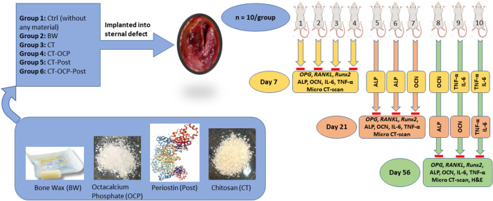
Methods: The pastes (new BW(s)) were prepared in the laboratory and examined by MTT, MIC, MBC, and degradability tests. Then, 60 adult male Wistar rats, divided into six equal groups including chitosan (CT), CT-octacalcium phosphate (OCP), CT-periostin (Post), CT-OCP-Post, Control (Ctrl), and BW, underwent sternotomy surgery. Once the surgeries were completed, the bone repair was assessed radiologically and thereafter clinically in vivo and in vitro using CT-scan, H&E, ELISA, and qRT-PCR.
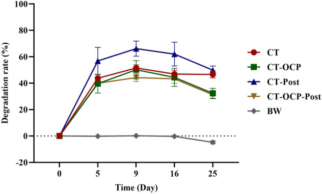
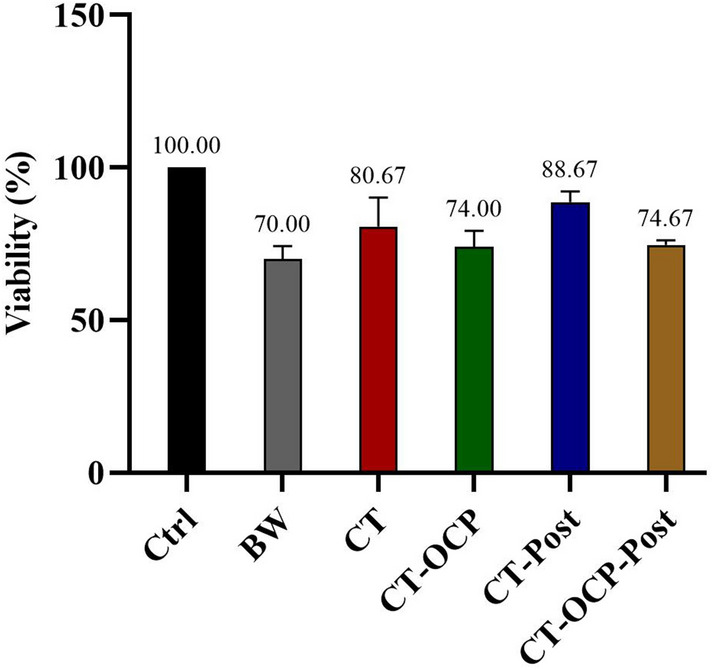
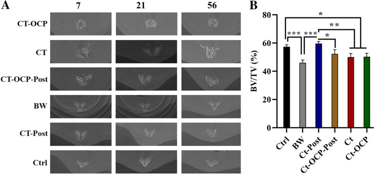
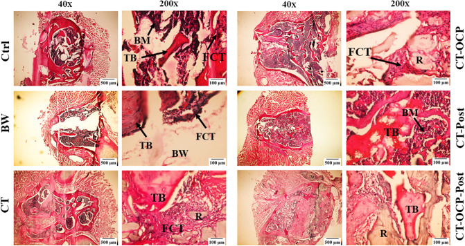
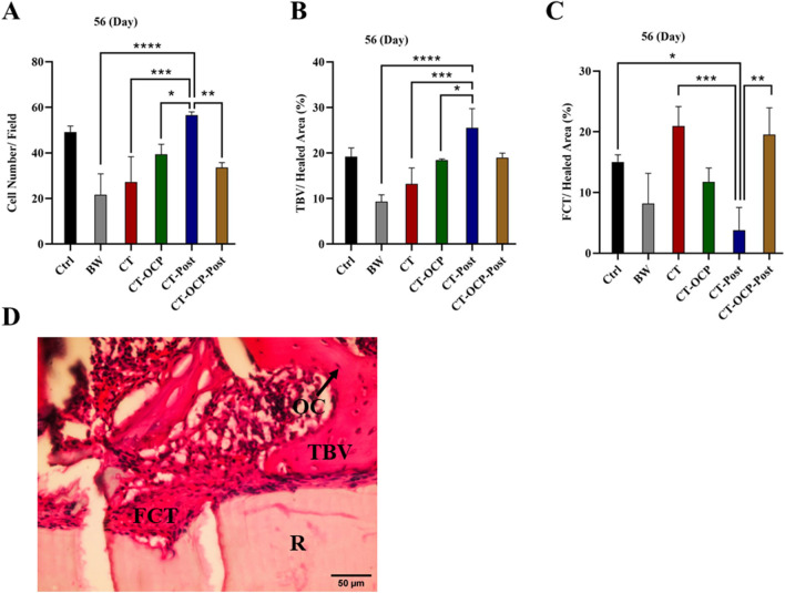
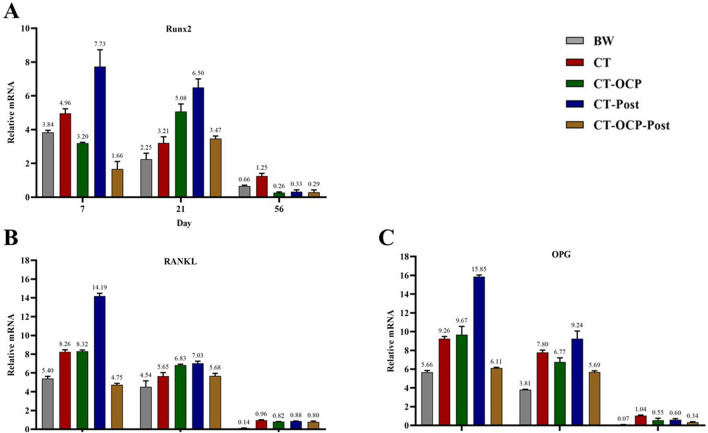
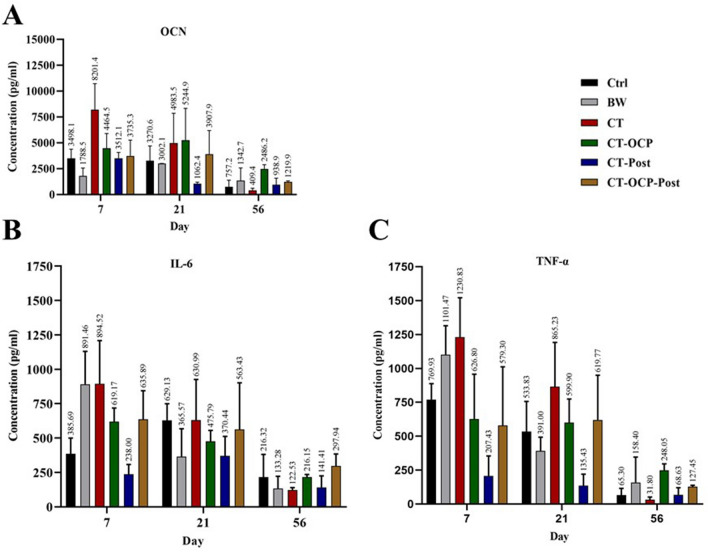
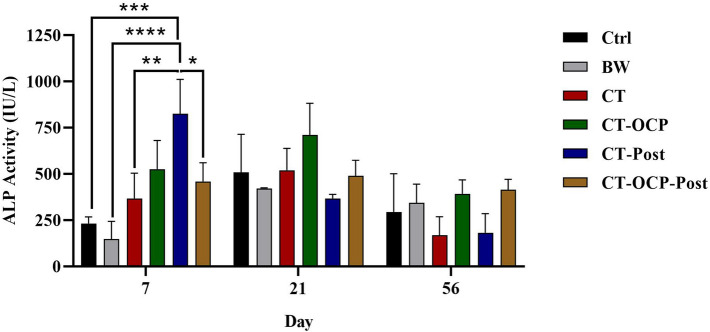
Results: All pastes displayed antibacterial properties and the CT-Post group had the highest cell viability compared to the control group. In contrast to the BW, CT-Post group demonstrated weight changes in the degradability test. In the CT-Post group, more number of osteocyte cells, high trabeculae percentage, and the least fibrous connective tissue were observed compared to other groups. Additionally, in comparison to the CT and Ctrl groups, higher alkaline phosphatase activity, as well as decreased level of serum tumor necrosis factor-α, interleukin-6, and OCN in the CT-Post group was evident. Finally, Runx2, OPG, and RANKL genes' expression was significantly higher in the CT-Post group than in other groups.
Conclusion: Our results provide insights into the desirability of pastes in terms of cellular viability, degradability, antibacterial properties, and surgical site restoration compared to the BW group. Besides, Periostin could enhance the osteogenic properties of bone tissue defect site.
Keywords: Bone healing; Chitosan; OPG and RANKL; Periostin; Runx2.
© 2022. The Korean Tissue Engineering and Regenerative Medicine Society.
Conflict of interest statement
The authors declare that they have no conflict of interest.
Figures
Similar articles
-
Comparison of bone wax and chitosan usage on post-sternotomy bone healing.Asian Cardiovasc Thorac Ann. 2021 Mar;29(3):203-207. doi: 10.1177/0218492320984097. Epub 2020 Dec 22. Asian Cardiovasc Thorac Ann. 2021. PMID: 33353370
-
[Mechanical properties and effect on osteodifferentiation of induced pluripotent stem cells of chitosan/whisker/calcium phosphate cement composite biomaterial].Zhongguo Xiu Fu Chong Jian Wai Ke Za Zhi. 2018 Jul 15;32(7):959-967. doi: 10.7507/1002-1892.201710028. Zhongguo Xiu Fu Chong Jian Wai Ke Za Zhi. 2018. PMID: 30129324 Free PMC article. Chinese.
-
Octacalcium Phosphate/Gelatin Composite (OCP/Gel) Enhances Bone Repair in a Critical-sized Transcortical Femoral Defect Rat Model.Clin Orthop Relat Res. 2022 Oct 1;480(10):2043-2055. doi: 10.1097/CORR.0000000000002257. Epub 2022 May 30. Clin Orthop Relat Res. 2022. PMID: 35638896 Free PMC article.
-
Anti-infective efficacy, cytocompatibility and biocompatibility of a 3D-printed osteoconductive composite scaffold functionalized with quaternized chitosan.Acta Biomater. 2016 Dec;46:112-128. doi: 10.1016/j.actbio.2016.09.035. Epub 2016 Sep 26. Acta Biomater. 2016. PMID: 27686039
-
Improved Healing by Adjuvant Osteoconductive Therapy Using a Novel Cotton-Like Hydroxyapatite Sheet After Median Sternotomy.Semin Thorac Cardiovasc Surg. 2020 Summer;32(2):244-252. doi: 10.1053/j.semtcvs.2019.10.018. Epub 2019 Nov 25. Semin Thorac Cardiovasc Surg. 2020. PMID: 31778787
Cited by
-
Immunotherapy targeting the obese white adipose tissue microenvironment: Focus on non-communicable diseases.Bioact Mater. 2024 Feb 19;35:461-476. doi: 10.1016/j.bioactmat.2024.01.027. eCollection 2024 May. Bioact Mater. 2024. PMID: 38404641 Free PMC article.
-
Impact of Polydeoxyribonucleotides on the Morphology, Viability, and Osteogenic Differentiation of Gingiva-Derived Stem Cell Spheroids.Medicina (Kaunas). 2024 Oct 1;60(10):1610. doi: 10.3390/medicina60101610. Medicina (Kaunas). 2024. PMID: 39459397 Free PMC article.
-
Exo-hydrogel therapy: a revolutionary approach to managing diabetic complications.J Nanobiotechnology. 2025 Aug 11;23(1):558. doi: 10.1186/s12951-025-03621-6. J Nanobiotechnology. 2025. PMID: 40790200 Free PMC article. Review.
-
Milk Consumption and Its Association with Dental Caries: Gender-Specific Insights from the Korea National Health and Nutrition Examination Survey (2013-2015).Medicina (Kaunas). 2024 Jun 11;60(6):967. doi: 10.3390/medicina60060967. Medicina (Kaunas). 2024. PMID: 38929584 Free PMC article.
-
Serum Periostin as a Novel Biomarker for Predicting 30-Day Major Adverse Cardiac Events After Off-Pump Coronary Artery Bypass Grafting.Ther Clin Risk Manag. 2025 Feb 18;21:161-176. doi: 10.2147/TCRM.S507435. eCollection 2025. Ther Clin Risk Manag. 2025. PMID: 39991461 Free PMC article.
References
-
- Oryan A, Alidadi S, Bigham-Sadegh A, Moshiri A, Kamali A. Effectiveness of tissue engineered chitosan-gelatin composite scaffold loaded with human platelet gel in regeneration of critical sized radial bone defect in rat. J Control Release. 2017;254:65–74. doi: 10.1016/j.jconrel.2017.03.040. - DOI - PubMed
MeSH terms
Substances
LinkOut - more resources
Full Text Sources