Review
. 2022 Jul;308(1):105-122.
doi: 10.1111/imr.13074. Epub 2022 Feb 23.

Extracellular vesicles and immune response during pregnancy: A balancing act

Affiliations
Review

Extracellular vesicles and immune response during pregnancy: A balancing act

Adrian E Morelli et al. Immunol Rev. 2022 Jul.

Abstract

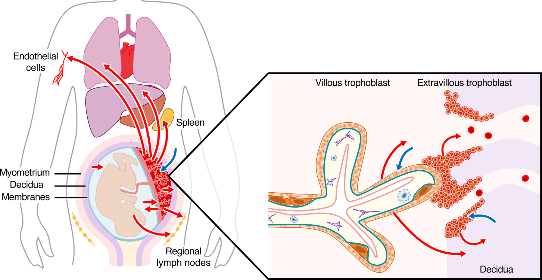
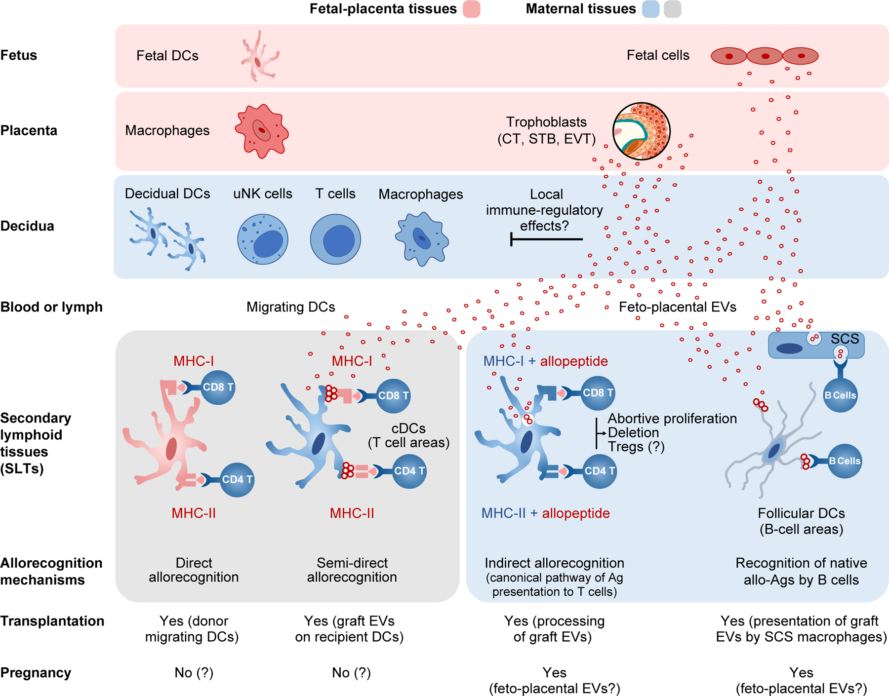
The mechanisms underlying maternal tolerance of the semi- or fully-allogeneic fetus are intensely investigated. Across gestation, feto-placental antigens interact with the maternal immune system locally within the trophoblast-decidual interface and distantly through shed cells and soluble molecules that interact with maternal secondary lymphoid tissues. The discovery of extracellular vesicles (EVs) as local or systemic carriers of antigens and immune-regulatory molecules has added a new dimension to our understanding of immune modulation prior to implantation, during trophoblast invasion, and throughout the course of pregnancy. New data on immune-regulatory molecules, located on EVs or within their cargo, suggest a role for EVs in negotiating immune tolerance during gestation. Lessons from the field of transplant immunology also shed light on possible interactions between feto-placentally derived EVs and maternal lymphoid tissues. These insights illuminate a potential role for EVs in major obstetrical disorders. This review provides updated information on intensely studied, pregnancy-related EVs, their cargo molecules, and patterns of fetal-placental-maternal trafficking, highlighting potential immune pathways that might underlie immune suppression or activation in gestational health and disease. Our summary also underscores the likely need to broaden the definition of the maternal-fetal interface to systemic maternal immune tissues that might interact with circulating EVs.

Keywords: extracellular vesicles; gestational immunology; pregnancy; tolerance.


Conflict of interest statement

CONFLICT OF INTEREST

Y. Sadovsky is a consultant at Illumina, Inc. The other authors report no conflicts.

Figures

Figure 1:
Figure 1:. Fetal-placental-maternal trafficking of EVs with potential immune functions in vivo.
The red arrows depict suggested feto-placental trafficking to the maternal decidua and uterine tissues, blood, secondary lymphoid tissues (uterus-draining lymph nodes, spleen), systemic endothelial cells, and distant organs. The blue arrows depict suggested trafficking routes of maternal EVs to feto-placental tissues. The inset depicts the routes of potential bi-directional EV passage at the maternal-placental-fetal interface.
Figure 2:
Figure 2:. Potential interactions between feto-placental EVs and maternal decidual leukocytes secondary lymphoid tissues.
Fetal tissues are noted in red shades, and maternal tissues in blue shades. The figure bottom compares mechanisms of allorecognition in the field of transplantation (gray shaded area) vs pregnancy, highlighting current knowledge. In mouse gestation, DCs develop relatively late, and a fairly low number of maternal leukocytes traffic to the fetal tissues. Thus, maternal T cells are unable to recognize allo-MHC molecules on fetal DCs mobilized to the mother’s SLTs (via the direct allorecognition pathway). Furthermore, because trophoblastic EVs do not express classic (highly polymorphic) MHC molecules on their surface, they are not recognized by directly alloreactive T cells on the surface of maternal APCs, when cross-dressed with the EVs (via the semi-direct allorecognition pathway, gray shaded area). Instead, feto-placental EVs may interact with maternal decidual uNK cells, macrophages, T cells, blood vessel cells, and decidual stromal cells, or traffic through peripheral blood or lymph to the maternal SLTs (spleen, uterus-draining lymph nodes), where the EVs are internalized and processed by maternal APCs (bottom blue shaded area.) In mice, fetus-derived antigenic peptides are presented within maternal MHC molecules by maternal APCs (likely conventional DCs) to T cells via the canonical pathway (also known, in transplantation, as the indirect pathway.) Feto-placental EVs could also be presented to maternal B cells by subcapsular sinus (SCS) macrophages in lymph nodes (or analogue macrophages in the spleen) of by follicular DCs located deep in B cell follicles.

Similar articles

Cited by

References

    1. Medawar PB. Some immunological and endocrinological problems raised by the evolution of viviparity in vertebrates. Symp Soc Exp Biol. 1953;7:320–338.
    1. Mor G, Aldo P, Alvero AB. The unique immunological and microbial aspects of pregnancy. Nat Rev Immunol. 2017;17(8):469–482. - PubMed
    1. Park JE, Jardine L, Gottgens B, Teichmann SA, Haniffa M. Prenatal development of human immunity. Science. 2020;368(6491):600–603. - PMC - PubMed
    1. Erlebacher A Immunology of the maternal-fetal interface. Annu Rev Immunol. 2013;31:387–411. - PubMed
    1. PrabhuDas M, Bonney E, Caron K, et al. Immune mechanisms at the maternal-fetal interface: Perspectives and challenges. Nat Immunol. 2015;16(4):328–334. - PMC - PubMed

Publication types