Review
. 2021 Dec 30;70(Suppl4):S597-S616.
doi: 10.33549/physiolres.934783.

Pancreatic stellate cells - rising stars in pancreatic pathologies

Affiliations
Review

Pancreatic stellate cells - rising stars in pancreatic pathologies

P Hrabák et al. Physiol Res. .

Abstract

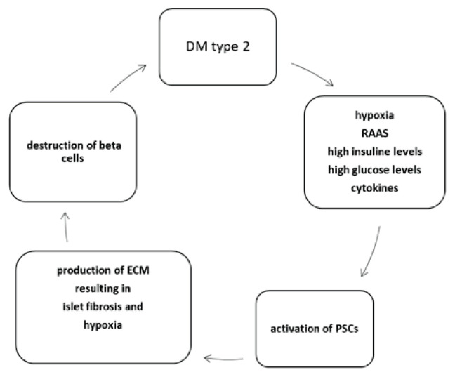
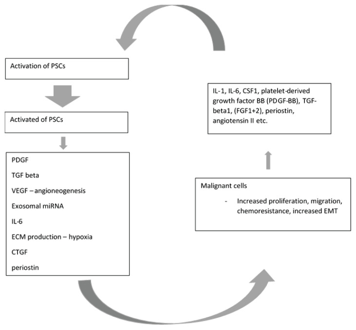
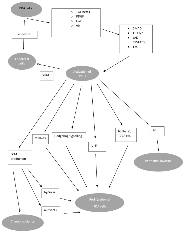
Pluripotent pancreatic stellate cells (PSCs) receive growing interest in past decades. Two types of PSCs are recognized -vitamin A accumulating quiescent PSCs and activated PSCs- the main producents of extracellular matrix in pancreatic tissue. PSCs plays important role in pathogenesis of pancreatic fibrosis in pancreatic cancer and chronic pancreatitis. PSCs are intensively studied as potential therapeutical target because of their important role in developing desmoplastic stroma in pancreatic cancer. There also exists evidence that PSC are involved in other pathologies like type-2 diabetes mellitus. This article brings brief characteristics of PSCs and recent advances in research of these cells.


Conflict of interest statement

Conflict of Interest

There is no conflict of interest.

Figures

Fig. 1
Fig. 1
Relationship between DM2 and PSCs. DM2 is a condition accompanied by environment rich in insulin and glucose within the pancreatic islets. High oxygen consumption by insulin producing beta cells results in hypoxia and oxidative stress. This, in combination with activation of angiotensin II pathway, leads to activation of PSCs. PSCs produce ECM and cytokines which results both directly and indirectly due to islet fibrosis in beta cells destruction. DM type 2 – Type 2 diabetes mellitus, PSCs – pancreatic stellate cells; RAAS – renin-angiotensin-aldosterone system; ECM – extracellular matrix. Modified from Kim et al. (2008).
Fig. 2
Fig. 2
Paracrine signalling via cytokines produced by malignant cells leads to PCSs activation. While activated, PSCs secrete factors, which increase ability of malignant cells to proliferate and migrate, increase metastatic potential and resistance to chemotherapeutics. Both malignant cells and activated PSCs produce factors which further activate PSCs. PDGF: platelet derived growth factor, VEGF – vascular endotelial growth factor, TGF – tumor growth factor; miRNA – microRNA; ECM - extracellular matrix; CTGF – connective tissue growth factor; EMT – epitelial-mesenchymal transformation; CSF1 – colony-stimulating factor 1; FGF – fibroblast growth factor. Modified from: Carter et al. (2021)
Fig. 3
Fig. 3
Crosstalk between malignant cells and PSCs. Factors produced by PSCs increase via several signalling pathways proliferation of malignant cells, perineural invasion, metastasing and chemoresistance. Both PSCs and malignant cells regulates angioneogenesis. Proliferation of malignant cells and hypoxia due to low blood supply increases activation of PSCs. Abbreviations: PDGF: platelet derived growth factor, VEGF – vascular endotelial growth factor, TGF – tumour growth factor; miRNA – microRNA; ECM – extracellular matrix; FGF – fibroblast growth factor; NGF – nerve growth factor; ERK extracellular signal-regulated kinases; JAK-Janus kinase; PDA – pancreatic ductal adenocarcinoma. Modified from: Bynigeri et al. (2017)

Similar articles

Cited by

References

    1. AOKI H, OHNISHI H, HAMA K, SHINOZAKI S, KITA H, YAMAMOTO H, OSAWA H, SATO K, TAMADA K, SUGANO K. Existence of autocrine loop between interleukin-6 and transforming growth factor-beta1 in activated rat pancreatic stellate cells. J Cell Biochem. 2006;99:221–228. doi: 10.1002/jcb.20906. - DOI - PubMed
    1. APTE MV, HABER PS, APPLEGATE TL, NORTON ID, MCCAUGHAN GW, KORSTEN MA, PIROLA RC, WILSON JS. Periacinar stellate shaped cells in rat pancreas: identification, isolation, and culture. Gut. 1998;43:128–133. doi: 10.1136/gut.43.1.128. - DOI - PMC - PubMed
    1. APTE MV, PHILLIPS PA, FAHMY RG, DARBY SJ, RODGERS SC, MCCAUGHAN GW, KORSTEN MA, PIROLA RC, NAIDOO D, WILSON JS. Does alcohol directly stimulate pancreatic fibrogenesis? Studies with rat pancreatic stellate cells. Gastroenterology. 2000;118:780–794. doi: 10.1016/s0016-5085(00)70148-x. - DOI - PubMed
    1. APTE MV, WILSON JS. Stellate cell activation in alcoholic pancreatitis. Pancreas. 2003;27:316–320. doi: 10.1097/00006676-200311000-00008. - DOI - PubMed
    1. ASAHINA K, TSAI SY, LI P, ISHII M, MAXSON RE, JR, SUCOV HM, TSUKAMOTO H. Mesenchymal origin of hepatic stellate cells, submesothelial cells, and perivascular mesenchymal cells during mouse liver development. epatology. 2009;49:998–1011. doi: 10.1002/hep.22721. - DOI - PMC - PubMed

MeSH terms