Recurrent Dermatofibrosarcoma Protuberance: A Case Report
- PMID: 35199742
- PMCID: PMC9124319
- DOI: 10.31729/jnma.7187
Recurrent Dermatofibrosarcoma Protuberance: A Case Report
Abstract
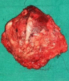
Dermatofibrosarcoma protuberance represents less than 0.1% of all tumors, treatment of which requires wide local excision (≥5cm) but recurrence is not rare. Here we present a 32-year male presented with a swelling of 15 x 6cm over the left lumbar region for which he underwent excision three years ago, the histopathological examination of the swelling, showed a malignant mesenchymal tumor and Immunohistochemistry features were suggestive of Dermatofibrosarcoma protuberance. After three years of interval, he again presented with complaints of swelling in the previously operated site for nine months and underwent excision of the mass with Split Thickness Skin Graft. Although the tumor was confined to the skin and subcutaneous tissue in the present case, the patient didn't undergo any adjuvant radiotherapy to avoid a possible relapse that would infiltrate deeper structures for the first time. Being a recurrent tumor, long-term follow-up is strongly recommended.
Keywords: adjuvant radiotherapy dermatofibrosarcoma; recurrence; skin grafting..
Conflict of interest statement
Figures
References
-
- Darier J, Ferrad M. [A case of elevated cutaneous fibrosarcoma that recurred 10 years later]. Ann Dermatol yphiligr. 1924;5:545–62. Japanese, English.
-
- Ghimire R, Subedi S, Sharma A, Bohara S. Anterior abdominal wall dermatofibrosarcoma protuberans: A case report. Journal of Society of Surgeons of Nepal. 2014;17(1):39–41. doi: 10.3126/jssn.v17i1.15180. - DOI
Publication types
MeSH terms
LinkOut - more resources
Full Text Sources
Medical


