

No evidence for pericardial restraint in the snapping turtle (Chelydra serpentina) following pharmacologically induced bradycardia at rest or during exercise
- PMID: 35200048
- PMCID: PMC9018006
- DOI: 10.1152/ajpregu.00004.2022
No evidence for pericardial restraint in the snapping turtle (Chelydra serpentina) following pharmacologically induced bradycardia at rest or during exercise
Abstract
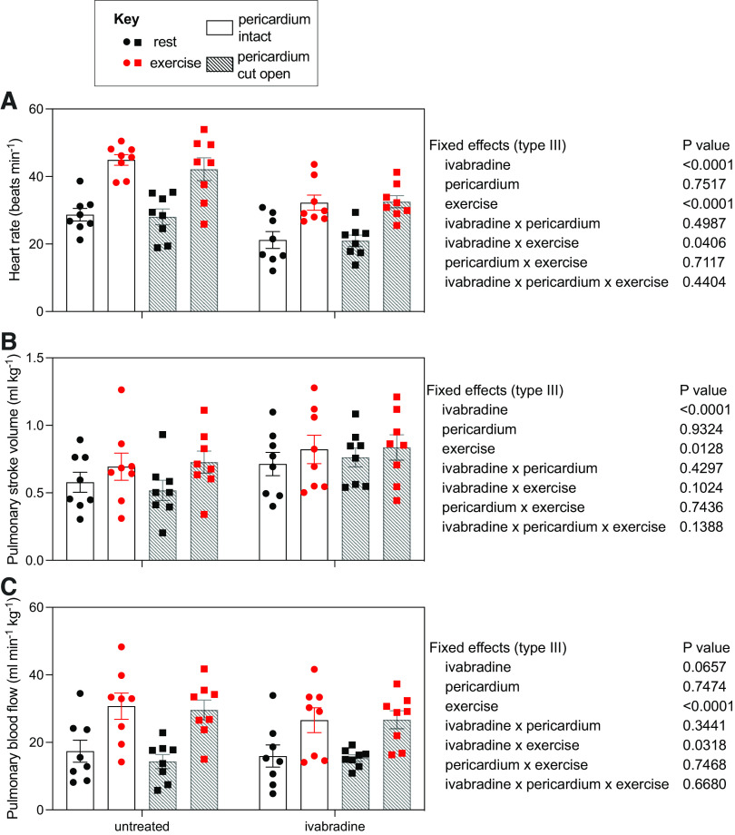
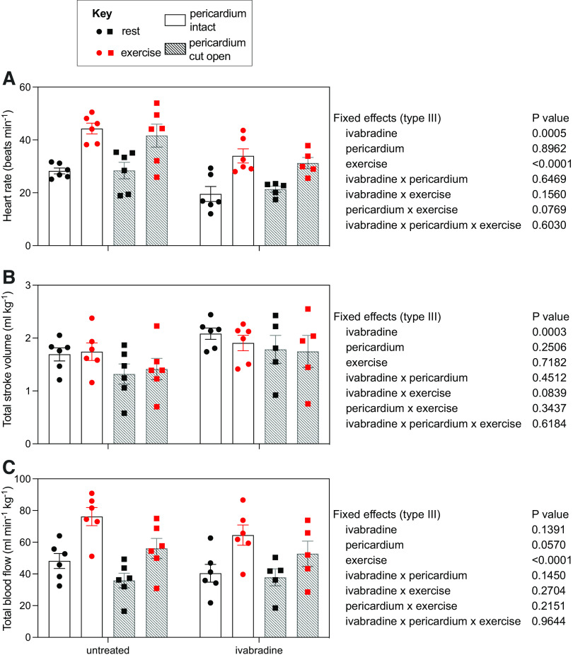
Most animals elevate cardiac output during exercise through a rise in heart rate (fH), whereas stroke volume (VS) remains relatively unchanged. Cardiac pacing reveals that elevating fH alone does not alter cardiac output, which is instead largely regulated by the peripheral vasculature. In terms of myocardial oxygen demand, an increase in fH is more costly than that which would incur if VS instead were to increase. We hypothesized that fH must increase because any substantial rise in VS would be constrained by the pericardium. To investigate this hypothesis, we explored the effects of pharmacologically induced bradycardia, with ivabradine treatment, on VS at rest and during exercise in the common snapping turtle (Chelydra serpentina) with intact or opened pericardium. We first showed that, in isolated myocardial preparations, ivabradine exerted a pronounced positive inotropic effect on atrial tissue but only minor effects on ventricle. Ivabradine reduced fH in vivo, such that exercise tachycardia was attenuated. Pulmonary and systemic VS rose in response to ivabradine. The rise in pulmonary VS largely compensated for the bradycardia at rest, leaving total pulmonary flow unchanged by ivabradine, although ivabradine reduced pulmonary blood flow during swimming (exercise × ivabradine interaction, P < 0.05). Although systemic VS increased, systemic blood flow was reduced by ivabradine both at rest and during exercise, despite ivabradine's potential to increase cardiac contractility. Opening the pericardium had no effect on fH, VS, or blood flows before or after ivabradine, indicating that the pericardium does not constrain VS in turtles, even during pharmacologically induced bradycardia.
Keywords: Testudines; activity; cardiovascular; ectotherm; reptile.
Conflict of interest statement
No conflicts of interest, financial or otherwise, are declared by the authors.
Figures


Similar articles
-
Embryonic hypoxia programmes postprandial cardiovascular function in adult common snapping turtles (Chelydra serpentina).J Exp Biol. 2017 Jul 15;220(Pt 14):2589-2597. doi: 10.1242/jeb.160549. Epub 2017 May 11. J Exp Biol. 2017. PMID: 28495871
-
Cardiovascular responses to putative chemoreceptor stimulation of embryonic common snapping turtles (Chelydra serpentina) chronically incubated in hypoxia (10% O2).Comp Biochem Physiol A Mol Integr Physiol. 2021 Sep;259:110977. doi: 10.1016/j.cbpa.2021.110977. Epub 2021 May 10. Comp Biochem Physiol A Mol Integr Physiol. 2021. PMID: 33984502
-
Maximum heart rate does not limit cardiac output at rest or during exercise in the American alligator ( Alligator mississippiensis).Am J Physiol Regul Integr Comp Physiol. 2018 Aug 1;315(2):R296-R302. doi: 10.1152/ajpregu.00027.2018. Epub 2018 Apr 25. Am J Physiol Regul Integr Comp Physiol. 2018. PMID: 29693431
-
Novel If current inhibitor ivabradine: safety considerations.Adv Cardiol. 2006;43:79-96. doi: 10.1159/000095430. Adv Cardiol. 2006. PMID: 16936474 Review.
-
Ivabradine: cardiovascular effects.Recent Pat Cardiovasc Drug Discov. 2009 Jan;4(1):61-6. doi: 10.2174/157489009787260016. Recent Pat Cardiovasc Drug Discov. 2009. PMID: 19149708 Review.
References
-
- Wang T, Joyce W, Hicks JW. Similitude in the cardiorespiratory responses to exercise across vertebrates. Curr Opin Physiol 10: 137–145, 2019. doi:10.1016/j.cophys.2019.05.007. - DOI
Publication types
MeSH terms
Substances
LinkOut - more resources
Full Text Sources
Miscellaneous