Effects of Prone Position on Lung Recruitment and Ventilation-Perfusion Matching in Patients With COVID-19 Acute Respiratory Distress Syndrome: A Combined CT Scan/Electrical Impedance Tomography Study
- PMID: 35200194
- PMCID: PMC9005091
- DOI: 10.1097/CCM.0000000000005450
Effects of Prone Position on Lung Recruitment and Ventilation-Perfusion Matching in Patients With COVID-19 Acute Respiratory Distress Syndrome: A Combined CT Scan/Electrical Impedance Tomography Study
Abstract
Objectives: Prone positioning allows to improve oxygenation and decrease mortality rate in COVID-19-associated acute respiratory distress syndrome (C-ARDS). However, the mechanisms leading to these effects are not fully understood. The aim of this study is to assess the physiologic effects of pronation by the means of CT scan and electrical impedance tomography (EIT).
Design: Experimental, physiologic study.
Setting: Patients were enrolled from October 2020 to March 2021 in an Italian dedicated COVID-19 ICU.
Patients: Twenty-one intubated patients with moderate or severe C-ARDS.
Interventions: First, patients were transported to the CT scan facility, and image acquisition was performed in prone, then supine position. Back to the ICU, gas exchange, respiratory mechanics, and ventilation and perfusion EIT-based analysis were provided toward the end of two 30 minutes steps (e.g., in supine, then prone position).
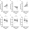
Measurements and main results: Prone position induced recruitment in the dorsal part of the lungs (12.5% ± 8.0%; p < 0.001 from baseline) and derecruitment in the ventral regions (-6.9% ± 5.2%; p < 0.001). These changes led to a global increase in recruitment (6.0% ± 6.7%; p < 0.001). Respiratory system compliance did not change with prone position (45 ± 15 vs 45 ± 18 mL/cm H2O in supine and prone position, respectively; p = 0.957) suggesting a decrease in atelectrauma. This hypothesis was supported by the decrease of a time-impedance curve concavity index designed as a surrogate for atelectrauma (1.41 ± 0.16 vs 1.30 ± 0.16; p = 0.001). Dead space measured by EIT was reduced in the ventral regions of the lungs, and the dead-space/shunt ratio decreased significantly (5.1 [2.3-23.4] vs 4.3 [0.7-6.8]; p = 0.035), showing an improvement in ventilation-perfusion matching.
Conclusions: Several changes are associated with prone position in C-ARDS: increased lung recruitment, decreased atelectrauma, and improved ventilation-perfusion matching. These physiologic effects may be associated with more protective ventilation.
Copyright © 2022 by the Society of Critical Care Medicine and Wolters Kluwer Health, Inc. All Rights Reserved.
Conflict of interest statement
Dr. Mauri received funding from the Department of Anesthesia, Critical Care and Emergency, Fondazione Istituto di Ricovero e Cura a Carattere Scientifico Ca’ Granda Ospedale Maggiore Policlinico, Milan, Italy (ricerca corrente 2020). Dr. Mauri’s institution received funding from Fisher and Paykel and Draeger; he received funding from Fisher and Paykel, Draeger, and BBraun. The remaining authors have disclosed that they do not have any potential conflicts of interest.
Figures


Comment in
-
Prone Position and COVID-19: Mechanisms and Effects.Crit Care Med. 2022 May 1;50(5):873-875. doi: 10.1097/CCM.0000000000005486. Epub 2022 Feb 7. Crit Care Med. 2022. PMID: 35120038 Free PMC article. No abstract available.
-
Prone Positioning for Patients With COVID-19-Associated Acute Respiratory Distress Syndrome.Crit Care Med. 2022 Oct 1;50(10):e772-e773. doi: 10.1097/CCM.0000000000005614. Epub 2022 Sep 12. Crit Care Med. 2022. PMID: 36106975 Free PMC article. No abstract available.
References
-
- Guérin C, Reignier J, Richard JC, et al. ; PROSEVA Study Group: Prone positioning in severe acute respiratory distress syndrome. N Engl J Med. 2013; 368:2159–2168 - PubMed

