

Intramedullary Nailing for Lower Limb Polyostotic Fibrous Dysplasia in Children: A Long-term Follow-up Study
- PMID: 35200214
- PMCID: PMC10810345
- DOI: 10.1097/BPO.0000000000002097
Intramedullary Nailing for Lower Limb Polyostotic Fibrous Dysplasia in Children: A Long-term Follow-up Study
Abstract
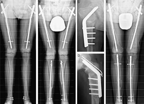
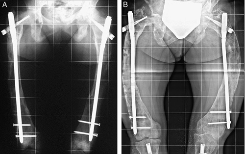
Background: In children, intramedullary nailing (IN) has been proposed as the best treatment when the femur and tibia are totally affected by fibrous dysplasia (FD). However, in younger children IN must be repeated to maintain stabilization of the affected skeletal segment during growth. We report the long-term results in a cohort of patients in whom more than two-thirds of cases had IN repeated during growth.
Methods: Twenty-nine femurs and 14 tibias totally affected by FD were treated by IN in 21 patients with polyostotic FD and McCune-Albright syndrome. Thirteen patients with 35 femoral and tibial deformities had a painful limp whereas 8 presented fractures. The patients had their first IN at a mean age of 9.26±2.68 years (range: 4 to 14 y). IN was repeated during growth in the younger patients, and all the patients underwent a mean of 2.13 femoral and 1.50 tibial IN per limb. The last IN was performed at a mean age of 16.42±1.95 years (range: 11 to 19 y). Titanium elastic nails and adult humeral nails were used in younger children, whereas adult femoral cervicodiaphyseal and interlocking tibial nails were used in older children and adolescents. At the latest follow-up, the patients were evaluated with a clinicoradiographic scale. All the data were statistically analyzed.
Results: The mean length of follow-up from the last IN was 6.47±3.10 years (range: 3 to 14 y), and the mean age of the patients at follow-up was 22.85±3.53 years (range: 14 to 29 y) when lower limbs were fully grown in all but 1 patient. Satisfactory long-term results were obtained in about 81% of our patients, while complications occurred in 32.5% of the 43 cases.
Conclusion: Lower limb IN-that was repeated in younger children during growth-provided satisfactory long-term results in most of our patients, with fracture and deformity prevention and pain control, regardless of the high rate of complications that mainly affected the femoral cases. Missing scheduled follow-ups was the main predictor of a poor result.
Level of evidence: Level IV-case series.
Copyright © 2022 Wolters Kluwer Health, Inc. All rights reserved.
Conflict of interest statement
The authors declare no conflicts of interest.
Figures


References
-
- DiCaprio MR, Enneking WF. Fibrous dysplasia: pathophysiology, evaluation, and treatment. J Bone Joint Surg Am. 2005;87:1848–1864. - PubMed
-
- Harris W, Dudley HR, Barry R. The natural history of fibrous dysplasia: an orthopaedic, pathological, and roentgenographic study. J Bone Joint Surg Am. 1962;44:207–233. - PubMed
-
- Ippolito E, Bray EW, Corsi A, et al. . Natural history and treatment of fibrous dysplasia of bone: a multicenter clinicopathologic study promoted by the European Pediatric Orthopaedic Society. J Pediatr Orthop B. 2003;12:155–177. - PubMed
-
- Ippolito E, Valentini MB, Lala R, et al. . Changing pattern of femoral deformity during growth in polyostotic fibrous dysplasia of the bone: an analysis of 46 cases. J Pediatr Orthop. 2016;36:488–493. - PubMed
-
- Leet AI, Chebli C, Kushner H, et al. . Fracture incidence in polyostotic fibrous dysplasia and the McCune-Albright syndrome. J Bone Miner Res. 2004;19:571–577. - PubMed
MeSH terms
LinkOut - more resources
Full Text Sources
Medical
Research Materials
