. 2022 Feb 9;9(2):67.
doi: 10.3390/bioengineering9020067.

An Immersible Microgripper for Pancreatic Islet and Organoid Research

Affiliations

An Immersible Microgripper for Pancreatic Islet and Organoid Research

Eike Früh et al. Bioengineering (Basel). .

Abstract

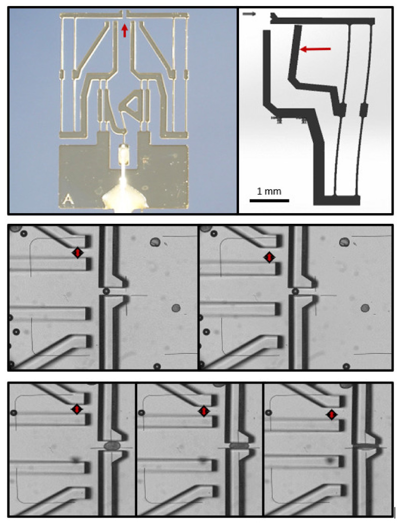
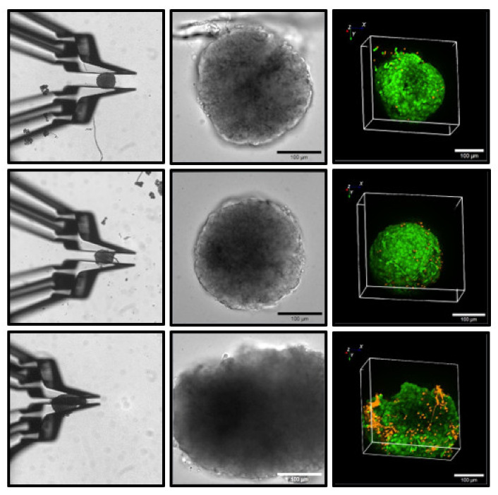
To improve the predictive value of in vitro experimentation, the use of 3D cell culture models, or organoids, is becoming increasingly popular. However, the current equipment of life science laboratories has been developed to deal with cell monolayers or cell suspensions. To handle 3D cell aggregates and organoids in a well-controlled manner, without causing structural damage or disturbing the function of interest, new instrumentation is needed. In particular, the precise and stable positioning in a cell bath with flow rates sufficient to characterize the kinetic responses to physiological or pharmacological stimuli can be a demanding task. Here, we present data that demonstrate that microgrippers are well suited to this task. The current version is able to work in aqueous solutions and was shown to position isolated pancreatic islets and 3D aggregates of insulin-secreting MIN6-cells. A stable hold required a gripping force of less than 30 μN and did not affect the cellular integrity. It was maintained even with high flow rates of the bath perfusion, and it was precise enough to permit the simultaneous microfluorimetric measurements and membrane potential measurements of the single cells within the islet through the use of patch-clamp electrodes.

Keywords: SU-8; cytosolic calcium; electrophysiology; microgripper; organoids; pancreatic islet.


Conflict of interest statement

The authors declare no conflict of interest. The funders had no role in the design of the study; in the collection, analyses, or interpretation of the data; in the writing of the manuscript; or in the decision to publish the results.

Figures

Figure 1
Figure 1
Design of a closed system for the pneumatic actuation of the gripper jaws. Increasing the air pressure protrudes the membrane at the tip of the tube whereby the jaws are set in motion. The left panel shows a 3D view of the entire setup. “1” denotes the membrane-sealed tube, and “2” denotes the microgripper. The right panel shows a detailed view of the gripper structure. The numbers denote the following: 1 = gear structure for generating a parallel movement of the gripper jaws; 2 = force measurement structures with optical markers; 3 = jaws; 4 = gear structure for the transmission of the membrane protrusion; 5 = actuator membrane (nonprotruding); and 6 = air pressure.
Figure 2
Figure 2
Measurement of the gripping force. (Upper row): overview of the entire microgripper with integrated force sensing (left), and schematic view of the principle of function (right). The arrow in the left panel points to the gap between the gripping jaws, and the one in the right panel indicates the deflected pointer. (Middle row): gripping of a noncompressible polyethylene microbead with minimal force (left), and with near maximal force (right). The microbead has a diameter of 100 µm, and the width of the gap increases from ca. 150 µm to ca. 200 µm, as indicated by the double-headed arrow. (Lower row): gripping with minimal force (left), reversible deformation by gripping (middle), and irreversible deformation by gripping (right). As indicated by the carets, only the force that irreversibly deforms the islet is sufficient to move the pointer by ca. 10%, which is equivalent to ca. 15 µm. This force can be estimated to correspond to ca. 30 µN.
Figure 3
Figure 3
Mechanical actuation with force transduction via Bowden cable. The upper image shows the entire setup. The numbers denote the following: 1 = microgripper; 2 = cannula; 3 = micrometer screw; 4 = stepper motor. The lower image shows the microgripper in close-up view. “1” denotes the site where the cannula is bonded to the microgripper base, and “2” denotes the site where the wire guided through the cannula is bonded to the force application point of the gripper gear.
Figure 4
Figure 4
Absence of cellular damage by gripping with minimal force (upper row), and with reversibly deforming force (middle row), and widespread occurrence of necrosis after irreversible deformation (lower row). The left column of images shows the aspect of the islets during gripping, and the middle column shows the aspect of the islets after gripping, both in transmitted light. The right column shows the 3D aspect of the respective islets as registered by confocal laser scanning fluorescence. Intact cells are marked by green fluorescence, and dead cells are marked by red fluorescence. Gripping without visible deformation remains without cellular damage in the islets. The force exerted in the experiment shown in the lower row can be estimated to be ca. 30 µN (compared with Figure 2).
Figure 5
Figure 5
Measurement of the plasma membrane potential of an MIN6 insulin-secreting cell within a pseudoislet. The (upper graph) shows the irregularly formed pseudoislet with the tip of the patch pipette approaching (left), and the tip attached to a single cell after seal formation (right). The (lower graph) shows the resulting registration. The pseudoislet was continuously perifused with a medium containing 1 mM of glucose. From 240 to 720 s, the medium additionally contained 500 µM of tolbutamide, and the potassium concentration was increased to 40 mM. Note the occurrence of action-potential spiking, which stops upon washout.
Figure 6
Figure 6
Measurement of the cytosolic Ca2+ concentration in a perifused islet concurrently with the measurement of the plasma membrane potential of a single islet cell. The (upper graph) shows the islet with the pipette tip attached to a single cell after seal formation (left), and the same islet emitting the fluorescence of the Ca2+ indicator, Fluo 4 (right). The (lower graph) shows the resulting registrations. The islet was continuously perifused with a medium containing 3 mM of glucose. From 400 to 1600 s, the medium additionally contained 500 µM of tolbutamide. Note the occurrence of action-potential spiking in the lower registration, which coincided with the increase in the cytosolic Ca2+ concentration (double-headed arrow). Upon washout, the spiking quickly ceased, as did the oscillatory pattern of the cytosolic Ca2+ concentration.

References

    1. Langer G. Implementation and Use of State-of-the-Art. In: Nielsch U., Fuhrmann U., Jaroch S., editors. New Approaches to Drug Discovery. Vol. 232. Springer; Cham/Basel, Switzerland: 2015. pp. 171–190. - DOI
    1. Cavero I., Guillon J.-M., Holzgrefe H.H. Human organotypic bioconstructs from organ-on-chip devices for human-predictive biological insights on drug candidates. Expert Opin. Drug Saf. 2019;18:651–677. doi: 10.1080/14740338.2019.1634689. - DOI - PubMed
    1. Garcés-Schröder M., Leester-Schädel M., Schulz M., Böl M., Dietzel A. Micro-Gripper: A new concept for a monolithic single-cell manipulation device. Sensors Actuators A Phys. 2015;236:130–139. doi: 10.1016/j.sna.2015.10.024. - DOI
    1. Gursky B., Bütefisch S., Leester-Schädel M., Li K., Matheis B., Dietzel A. A Disposable Pneumatic Microgripper for Cell Manipulation with Image-Based Force Sensing. Micromachines. 2019;10:707. doi: 10.3390/mi10100707. - DOI - PMC - PubMed
    1. Saba R., Iqbal S., Shakoor R.I., Saleem M.M., Bazaz S.A. Design and analysis of four-jaws microgripper with integrated thermal actuator and force sensor for biomedical applications. Rev. Sci. Instruments. 2021;92:045007. doi: 10.1063/5.0032404. - DOI - PubMed

LinkOut - more resources