Review
. 2022 Jan 28;8(2):82.
doi: 10.3390/gels8020082.

Drug Delivery Challenges and Current Progress in Nanocarrier-Based Ocular Therapeutic System

Affiliations
Review

Drug Delivery Challenges and Current Progress in Nanocarrier-Based Ocular Therapeutic System

Md Habban Akhter et al. Gels. .

Abstract

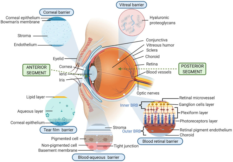
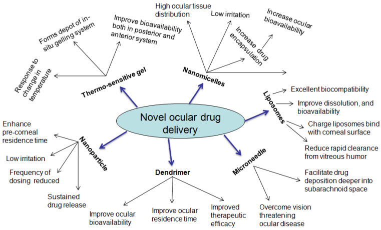
Drug instillation via a topical route is preferred since it is desirable and convenient due to the noninvasive and easy drug access to different segments of the eye for the treatment of ocular ailments. The low dose, rapid onset of action, low or no toxicity to the local tissues, and constrained systemic outreach are more prevalent in this route. The majority of ophthalmic preparations in the market are available as conventional eye drops, which rendered <5% of a drug instilled in the eye. The poor drug availability in ocular tissue may be attributed to the physiological barriers associated with the cornea, conjunctiva, lachrymal drainage, tear turnover, blood-retinal barrier, enzymatic drug degradation, and reflex action, thus impeding deeper drug penetration in the ocular cavity, including the posterior segment. The static barriers in the eye are composed of the sclera, cornea, retina, and blood-retinal barrier, whereas the dynamic barriers, referred to as the conjunctival and choroidal blood flow, tear dilution, and lymphatic clearance, critically impact the bioavailability of drugs. To circumvent such barriers, the rational design of the ocular therapeutic system indeed required enriching the drug holding time and the deeper permeation of the drug, which overall improve the bioavailability of the drug in the ocular tissue. This review provides a brief insight into the structural components of the eye as well as the therapeutic challenges and current developments in the arena of the ocular therapeutic system, based on novel drug delivery systems such as nanomicelles, nanoparticles (NPs), nanosuspensions, liposomes, in situ gel, dendrimers, contact lenses, implants, and microneedles. These nanotechnology platforms generously evolved to overwhelm the troubles associated with the physiological barriers in the ocular route. The controlled-drug-formulation-based strategic approach has considerable potential to enrich drug concentration in a specific area of the eye.

Keywords: drug delivery; hydrogel; implant; microneedle; nanomicelles; nanoparticles; ocular therapeutic system.


Conflict of interest statement

The authors declare no conflict of interest.

Figures

Figure 1
Figure 1
The anatomy of ocular system representing anterior (A) and posterior segments (B). The anterior segment includes conjunctiva, ciliary body, iris, pupil, anterior chamber, cornea, and lens. The posterior segment consists of sclera, choroid, retina, macula, and optic nerve. Modified from ref. [8]; permission under a creative common license (CC BY-NC-ND 4.0).
Figure 2
Figure 2
Drug delivery barriers in ocular route. The barriers may be from the anterior segment including corneal barriers, e.g., corneal epithelium, tear film, conjunctiva, and blood–aqueous barrier. The posterior segment barrier may be due to the blood–retinal barriers comprising retinal vessels, ganglion cells, pigment cells, retinal endothelium, and the vitreous barrier. These barriers overall reduced drug availability in the ocular tissues of the posterior segment. Permission received under Creative Commons Attribution 4.0 International License [17].
Figure 3
Figure 3
Conventional mode of ocular therapeutic system.
Figure 4
Figure 4
Nanocarriers employed in ocular therapeutic systems.
Figure 5
Figure 5
Diagram showing the route of PEGylated ME entry into the posterior segment of eye (A) and different parts of retina of eye (B). To overcome the cellular barriers, topical PEGylated ME may cross the membrane barriers viz. cornea, conjunctiva, and sclera, thereby preventing opsonization and improving circulation in lachrymal fluid and vitreous humor. Modified from ref. [2]; permission granted from ACS publishers (https://pubs.acs.org/doi/10.1021/acsomega.9b04244, (accessed on 20 December 2021)).
Figure 6
Figure 6
(I) Culture plate’s sterility test on incubation with (A) saline solution, (B) positive control, (C) PEGylated ME, and (D) normal ME; (II) tonicity evaluation, RBCs treated with (A) saline solution, (B) hypotonic solution, (C) hypertonic solution, (D) normal ME, and (E) microscopy of PEGylated ME; (III) hematoxylin-and-eosin-stained corneal sections treated with (A) saline solution, (B) normal ME, and (C) PEGylated ME observed under a microscope; (IV) corneal hydration test. Images captured after 3 h of hen’s egg membrane treated with (A) saline solution, (B) NaOH solution, (C) normal ME, and (D) PEGylated ME. Modified from ref. [2] (https://pubs.acs.org/doi/10.1021/acsomega.9b04244, (accessed on 20 December 2021)).
Figure 7
Figure 7
Novel therapeutic strategy in ocular drug delivery.
Figure 8
Figure 8
Investigation of liposomal effectiveness in ocular drug delivery: (a) the fluorescence images of different formulations with Coumarin (Cou) using cell analyzer. The cellular uptake after 24 h of human corneal epithelial cells (HCECs) (scale bar = 300 μm); (b) formulations intake count; (c) the Nile red-stained formulation distribution images captured in cornea; the corneal endothelium indicated by arrow (scale bar = 50 μm); (d) in vivo pharmacokinetic parameters after topical instillation of different formulations. Permission under Commons Attribution 4.0 International License [95]. (http://creativecommons.org/licenses/by/4.0/, (accessed on 15 November 2021)).
Figure 9
Figure 9
Effective drug delivery of liposomes into posterior ocular segment. Protection against photo-oxidative retinal damage: (a) the b-wave amplitude alteration following topical administration of different formulations for up to 14days; (b) retinographic images of various formulations treatment; (c) the protection efficacy of various hematoxylin-and-eosin (H and E)-stained formulations in the retina (scale bar = 20 μm); (d) images of in vitro anti-ROS efficacy taken with a long-term real-time dynamic live cell imaging analyzer: (e) ROS levels of various formulations. Data are expressed as mean ± SD (n = 3). * p < 0.05, ## p < 0.01, ** p < 0.01, *** p < 0.001. Permission under Commons Attribution 4.0 International License [95]. (http://creativecommons.org/licenses/by/4.0/, (accessed on 15 November 2021)).
Figure 10
Figure 10
(a,b) Ocular irritation studies: (a) typical histological image of formulation, P-CBLs after instillation for 14 successive days (scale bar = 20 μm); (b) ocular surface examination using a silt lamp and camera, post staining with 0.5 % sodium fluorescein. The ocular irritation studies manifested no injuries or abnormalities in either part of the cornea, conjunctiva, or iris of the eye (a). The 0.5% sodium fluorescein stained ocular surface observed under a silt lamp and camera found no edema or injuries, which further substantiated the safe and protective nature of P-CBLs (b). Permission under Commons Attribution 4.0 International License [95]. (http://creativecommons.org/licenses/by/4.0/, (accessed on 15 November 2021)).

References

    1. Gholizadeh S., Wang Z., Chen X., Dana R., Annabi N. Advanced nanodelivery platforms for topical ophthalmic drug delivery. Drug Discov. Today. 2021;26:1437–1449. doi: 10.1016/j.drudis.2021.02.027. - DOI - PubMed
    1. Nayak K., Misra M. Triamcinolone Acetonide-Loaded PEGylated Microemulsion for the Posterior Segment of Eye. ACS Omega. 2020;5:7928–7939. doi: 10.1021/acsomega.9b04244. - DOI - PMC - PubMed
    1. Bachu R.D., Chowdhury P., Al-Saedi Z.H.F., Karla P.K., Boddu S.H.S. Ocular Drug Delivery Barriers—Role of Nanocarriers in the Treatment of Anterior Segment Ocular Diseases. Pharmaceutics. 2018;10:28. doi: 10.3390/pharmaceutics10010028. - DOI - PMC - PubMed
    1. Almeida H., Amaral M.H., Lobao P., Lobo J.M.S. In situ gelling systems: A strategy to improve the bioavailability of ophthalmic pharmaceutical formulations. Drug Discov. Today. 2014;19:400–412. doi: 10.1016/j.drudis.2013.10.001. - DOI - PubMed
    1. Gorantla S., Rapalli V.K., Waghule T., Singh P.P., Dubey S.K., Saha R.N., Singhvi G. Nanocarriers for ocular drug delivery: Current status and translational opportunity. RSC Adv. 2020;10:27835–27855. doi: 10.1039/D0RA04971A. - DOI - PMC - PubMed

LinkOut - more resources