. 2022 Feb 16;5(1):19.
doi: 10.3390/mps5010019.

Quantitative Studies on the Interaction between Saposin-like Proteins and Synthetic Lipid Membranes

Affiliations

Quantitative Studies on the Interaction between Saposin-like Proteins and Synthetic Lipid Membranes

Suzanne I Sandin et al. Methods Protoc. .

Abstract

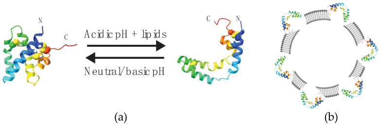
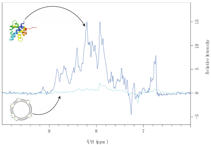
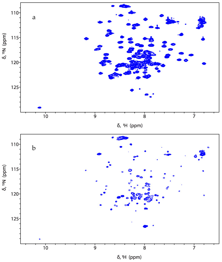
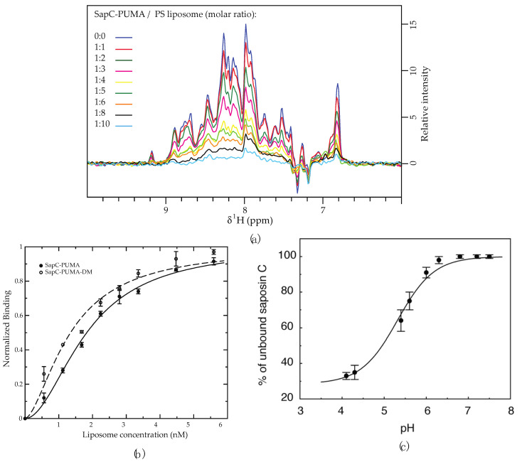
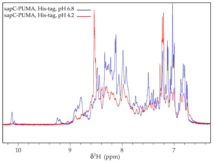
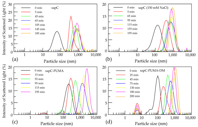
Members of the saposin-fold protein family and related proteins sharing a similar fold (saposin-like proteins; SAPLIP) are peripheral-membrane binding proteins that perform essential cellular functions. Saposins and SAPLIPs are abundant in both plant and animal kingdoms, and peripherally bind to lipid membranes to play important roles in lipid transfer and hydrolysis, defense mechanisms, surfactant stabilization, and cell proliferation. However, quantitative studies on the interaction between proteins and membranes are challenging due to the different nature of the two components in relation to size, structure, chemical composition, and polarity. Using liposomes and the saposin-fold member saposin C (sapC) as model systems, we describe here a method to apply solution NMR and dynamic light scattering to study the interaction between SAPLIPs and synthetic membranes at the quantitative level. Specifically, we prove with NMR that sapC binds reversibly to the synthetic membrane in a pH-controlled manner and show the dynamic nature of its fusogenic properties with dynamic light scattering. The method can be used to infer the optimal pH for membrane binding and to determine an apparent dissociation constant (KDapp) for protein-liposome interaction. We propose that these experiments can be applied to other proteins sharing the saposin fold.

Keywords: NMR; dynamic light scattering; liposomes; membrane fusion; protein-membrane interactions; saposin C; saposin-like proteins.


Conflict of interest statement

The authors declare no conflict of interest.

Figures

Figure 1
Figure 1
SapC undergoes a pH-dependent conformational change upon peripheral binding to liposomes. (a) Ribbon diagrams of soluble (closed form, left) and membrane-bound (V-shape form, right) sapC conformations determined by high-resolution NMR [2,3]. Arrows indicate reversibility with pH in binding and conformational change. (b) SapC in open conformation binds to the liposome surface.
Figure 2
Figure 2
Schematic representation of the effect of liposome binding on NMR signal intensity. NMR signal intensity from soluble sapC in the absence of liposomes (dark blue) and residual intensity in the presence of liposomes resulting from ~5% sapC free in solution (light blue).
Figure 3
Figure 3
SapC-PUMA binding to liposomes results in decreased signal intensity. 2D [1H-15N]-SOFAST-HMQC [18] spectra of sapC-PUMA at pH 6.0 in the absence of liposomes (a) and at 1:5 molar ratio of sapC-PUMA:PS lipids (b).
Figure 4
Figure 4
The binding of sapC constructs to liposomes results in NMR signal intensity decrease. (a) Signal intensity of sapC-PUMA amide 1H of 1D projections from 2D [1H-15N]-SOFAST-HMQC [18] decreases with increasing lipid concentration. In the absence of liposomes (dark blue) the signal intensity is normalized to 1. (b) The binding affinity of sapC-PUMA constructs to liposomes can be determined by fitting to the Hill equation. Experiments are conducted in duplicate. Bars indicate experimental errors. (c) SapC binding to PC:PS liposomes increases under acidic conditions [2]. (a,b): Reprinted from Pharmaceutics, 13 (2021) 583 (c): Reprinted from Biochemistry, 42 (2003) 14,729−14,740. Published 2003 American Chemical Society.
Figure 5
Figure 5
SapC-PUMA aggregates at acidic pH when the His-tag is not removed. 1D 1H-NMR spectrum of sapC-PUMA at 6.8 and 4.2 in the presence of His-tag. Reprinted from Pharmaceutics, 13 (2021) 583.
Figure 6
Figure 6
SapC constructs induce liposome fusion. Initial liposome size is shown in black at ~100–200 nm. Peaks appearing below 10 nm correspond to unbound protein. The following conditions were tested: sapC in water pH 4.3 (a), sapC with 150 mM NaCl pH 4.3 (b), sapC-PUMA in water pH 5.3 (c), and sapC-PUMA-DM in water pH 5.3 (d). Reprinted from Pharmaceutics, 13 (2021) 583.

Similar articles

Cited by

References

    1. Darmoise A., Maschmeyer P., Winau F. The Immunological Functions of Saposins. Adv. Immunol. 2010;105:25–62. - PMC - PubMed
    1. de Alba E., Weiler S., Tjandra N. Solution Structure of Human Saposin C: pH-Dependent Interaction with Phospholipid Vesicles. Biochemistry. 2003;42:14729–14740. doi: 10.1021/bi0301338. - DOI - PubMed
    1. Hawkins C.A., Alba E.D., Tjandra N. Solution Structure of Human Saposin C in a Detergent Environment. J. Mol. Biol. 2005;346:1381–1392. doi: 10.1016/j.jmb.2004.12.045. - DOI - PubMed
    1. Bruhn H. A Short Guided Tour through Functional and Structural Features of Saposin-Like Proteins. Biochem. J. 2005;389:249–257. doi: 10.1042/BJ20050051. - DOI - PMC - PubMed
    1. Falco A., Medina-Gali R.M., Poveda J.A., Bello-Perez M., Novoa B., Encinar J.A. Antiviral Activity of a Turbot (Scophthalmus Maximus) NK-Lysin Peptide by Inhibition of Low-pH Virus-Induced Membrane Fusion. Mar. Drugs. 2019;17:87. doi: 10.3390/md17020087. - DOI - PMC - PubMed

LinkOut - more resources