Review
. 2022 Jan 18;29(2):411-422.
doi: 10.3390/curroncol29020037.

Case Report of Small Cell Carcinoma of the Ovary, Hypercalcemic Type (Ovarian Rhabdoid Tumor) with SMARCB1 Mutation: A Literature Review of a Rare and Aggressive Condition

Affiliations
Review

Case Report of Small Cell Carcinoma of the Ovary, Hypercalcemic Type (Ovarian Rhabdoid Tumor) with SMARCB1 Mutation: A Literature Review of a Rare and Aggressive Condition

Maria Fernanda Evangelista Simões et al. Curr Oncol. .

Abstract

Small cell carcinoma of the ovary, hypercalcemic type (SCCOHT) is a rare and aggressive condition that is associated with the SMARCA4 mutation and has a dismal prognosis. It is generally diagnosed in young women. Here, we report a case of a young woman with SCCOHT harboring a rare molecular finding with a highly aggressive biological behavior. The patient had a somatic SMARCB1 mutation instead of an expected SMARCA4 alteration. Even though the patient was treated with high-dose chemotherapy followed by stem cell transplantation, she evolved with disease progression and died 11 months after her first symptoms appeared. We present a literature review of this rare disease and discuss the findings in the present patient in comparison to expected molecular alterations and options for SCCOHT treatment.

Keywords: SMARCB1 mutation; hypercalcemic type; ovarian cancer; small cell carcinoma of the ovary.


Conflict of interest statement

The authors declare no conflict of interest.

Figures

Figure 1
Figure 1
Computed tomography study in the portal phase showing a heterogeneous mass in the pelvic region (orange arrow), displacement of the intestinal loops to the right (blue arrow), and compression over the inferior vena cava (green arrow).
Figure 2
Figure 2
Magnetic resonance study in T1 weighted sequence post gadolinium showing a heterogeneous mass in the pelvic region (orange arrows).
Figure 3
Figure 3
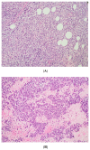
(A) The tumor cells are predominantly monomorphic and loosely cohesive with scant cytoplasm. (B) Small nests and individual tumor cells are separated by a fibrotic stroma. (C) A small cell component with nested growth and focal spindle morphology is juxtaposed to large cells with abundant eosinophilic cytoplasm. (D) The presence of areas of a more rhabdoid pattern are seen.
Figure 3
Figure 3
(A) The tumor cells are predominantly monomorphic and loosely cohesive with scant cytoplasm. (B) Small nests and individual tumor cells are separated by a fibrotic stroma. (C) A small cell component with nested growth and focal spindle morphology is juxtaposed to large cells with abundant eosinophilic cytoplasm. (D) The presence of areas of a more rhabdoid pattern are seen.
Figure 4
Figure 4
The pathology analysis of the surgical specimen, 100×. Sheets of tumor cells are interrupted by follicle-like spaces (H&E, 100×).
Figure 5
Figure 5
The pathology analysis of the surgical specimen. The stain INI-1—loss of expression (IHC, 100×).
Figure 6
Figure 6
(A) The pathology analysis of the surgical specimen. The SMARCA4 expression is retained (IHC, 100×). (B) The pathology analysis of the surgical specimen. Lack of nuclear expression of SMARCA2 is noted (left) with the internal control of the SMARCA2 in the ovarian stroma (right). (IHC, 100×).
Figure 7
Figure 7
The computed tomography study in the portal phase showing a lymph node in the left iliac chain with a rounded aspect and a necrotic center (red arrow), suggestive of secondary neoplastic involvement (relapse).
Figure 8
Figure 8
The computed tomography study in the portal phase showing a heterogeneous lesion in the pelvic region with cystic areas in between (orange arrows).
Figure 9
Figure 9
The computed tomography study in the portal phase demonstrates liver metastases (green arrows) and abdominal metastases (purple arrows).
Figure 10
Figure 10
The computed tomography study in the portal phase showing the dilation of slender loops (yellow arrows) by distal extrinsic compression (new pelvic lesion).

Similar articles

Cited by

References

    1. Tischkowitz N., Huang S., Banerjee S., Hague J., Hendricks W.P., Huntsman D.G., Lang J.D., Orlando K.A., Oza A.M., Pautier P., et al. Small-Cell Carcinoma of the Ovary, Hypercalcemic Type–Genetics, New Treatment Targets, and Current Management Guideline. Clin. Cancer Res. 2020;26:3908–3917. doi: 10.1158/1078-0432.CCR-19-3797. - DOI - PMC - PubMed
    1. Young R.H., Goodman A., Penson R.T., Russell A.H., Uppot R.N., Tambouret R.H. Case records of the Massachusetts General Hospital. Case 8-2010. A 22-year-old woman with hypercalcemia and a pelvic mass. N. Engl. J. Med. 2010;362:1031–2040. doi: 10.1056/NEJMcpc1000272. - DOI - PubMed
    1. Young R.H., Oliva E., Scully R.E. Small cell carcinoma of the ovary, hypercalcemic type. A clinicopathological analysis of 150 cases. Am. J. Surg. Pathol. 1994;18:1102–1116. doi: 10.1097/00000478-199411000-00004. - DOI - PubMed
    1. Ramos P., Karnezis A.N., Craig D.E., Sekulic A., Russell M.L., Hendricks W.P., Corneveaux J.J., Barrett M.T., Shumansky K., Yang Y., et al. Small cell carcinoma of the ovary, hypercalcemic type, displays frequent inactivating germline and somatic mutations in SMARCA4. Nat. Genet. 2014;46:427–429. doi: 10.1038/ng.2928. - DOI - PMC - PubMed
    1. Seidman J.D. Small cell carcinoma of the ovary of the hypercalcemic type: p53 protein accumulation and clinicopathologic features. Gynecol. Oncol. 1995;59:283–287. doi: 10.1006/gyno.1995.0023. - DOI - PubMed