. 2022 Feb 11;29(2):1018-1028.
doi: 10.3390/curroncol29020087.

Assessing Choroidal Nevi, Melanomas and Indeterminate Melanocytic Lesions Using Multimodal Imaging-A Retrospective Chart Review

Affiliations

Assessing Choroidal Nevi, Melanomas and Indeterminate Melanocytic Lesions Using Multimodal Imaging-A Retrospective Chart Review

Fredy Geiger et al. Curr Oncol. .

Abstract

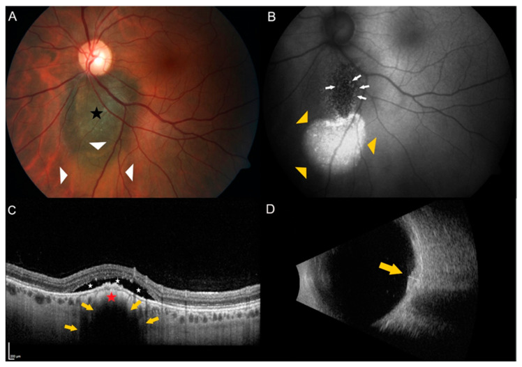
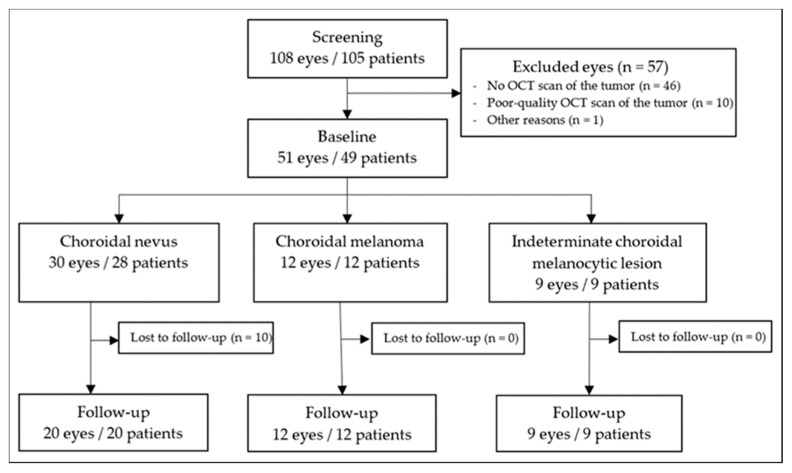
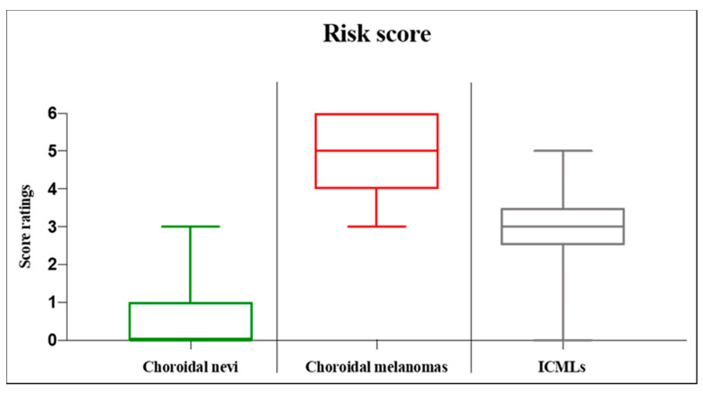
Using multimodal imaging, the literature proposed the following risk factors for choroidal nevus growth into melanoma: increased tumor thickness, subretinal fluid, decreased visual acuity, presence of orange pigment, ultrasound acoustic hollowness, and increased tumor diameter. This study investigated the presence of the mentioned risk factors in choroidal nevi, choroidal melanomas, and indeterminate choroidal melanocytic lesions. This retrospective, single-center chart review assessed choroidal melanocytic tumors with multimodal imaging. We defined our primary outcome as the cumulative presence of mentioned risk factors. Further, we evaluated various optical coherence tomography (OCT), ultrasound, and autofluorescence findings. We analyzed 51 tumors from 49 patients during the period from April 2008 to June 2021. The median (IQR) age was 64.0 (56.0 to 70.5) years, with 23 of 49 (46.9%) patients being female. The follow-up time for all tumors was median (IQR) 25.0 (12.0 to 39.0) months. The choroidal nevi had a median (range) risk score of 0.0 (0.0 to 3.0), and the choroidal melanoma of 5.0 (3.0 to 6.0), with statistically significant different ratings (p < 0.001). Multimodal imaging creates a score that may help to distinguish choroidal nevi from choroidal melanomas objectively.

Keywords: choroidal nevus; choroidal tumors; melanoma; multimodal imaging; ophthalmic oncology; optical coherence tomography.


Conflict of interest statement

The authors declare no conflict of interest.

Figures

Figure 1
Figure 1
Example of a juxtapapillary indeterminate choroidal melanocytic lesion showing two risk factors. (A) Color fundus photography of the left eye of a 35-year old female patient showing a pigmented choroidal lesion (black star) adjacent to the optic disc. Please note the presence of subretinal fluid over and inferior to the lesion (white triangles). (B) Autofluorescence imaging highlights the subretinal fluid (yellow triangles) and shows the presence of orange pigment (white arrows) over the lesion. (C) Enhanced depth spectral-domain optical coherence tomography (EDI-OCT) of the dome-shaped tumor (red star) shows choriocapillaris compression and choroidal shadowing (yellow arrows) as well as subretinal fluid (white stars) over the tumor. (D) B-scan ultrasonography shows a dense, dome-shaped lesion (yellow arrow) measuring 1.5 mm in thickness.
Figure 2
Figure 2
Flowchart illustrating study inclusion and exclusion. OCT = optical coherence tomography.
Figure 3
Figure 3
Boxplots displaying the analysis of the risk score for choroidal melanocytic lesions. The score consists of the following risk factors proposed by Shields et al. regarding choroidal nevus transformation to melanoma [14]: lesion thickness, subretinal fluid, Snellen visual acuity, orange pigment, ultrasound acoustic hollowness, and tumor diameter. A cumulative score between 0 and 6 points is possible, with the highest score reflecting all risk factors present. The box displays the first and third quartiles, and the line within the median value. The whiskers represent the minimum and maximum value. ICML = indeterminate choroidal melanocytic lesion.

References

    1. Yanoff M., Zimmerman L.E. Histogenesis of malignant melanomas of the uvea. II. Relationship of uveal nevi to malignant melanomas. Cancer. 1967;20:493–507. doi: 10.1002/1097-0142(1967)20:4<493::AID-CNCR2820200406>3.0.CO;2-U. - DOI - PubMed
    1. Arnesen K., Nornes M. Malignant melanoma of the choroid as related to coexistent benign nevus. Acta Ophthalmol. 1975;53:139–152. doi: 10.1111/j.1755-3768.1975.tb01148.x. - DOI - PubMed
    1. Prager A.J., Habib L.A., Busam K.J., Marr B.P. Two Uveal Melanomas in One Eye: A Choroidal Nevus Giving Rise to a Melanoma in an Eye with a Separate Large Choroidal Melanoma. Ocul. Oncol. Pathol. 2018;4:355–358. doi: 10.1159/000486682. - DOI - PMC - PubMed
    1. You Q.S., Xu L., Jonas J.B., Wang S., Yang H. Change in choroidal nevi during a 5-year follow-up study: The Beijing Eye Study. Br. J. Ophthalmol. 2010;94:575–578. doi: 10.1136/bjo.2009.165720. - DOI - PubMed
    1. Callaway N.F., Mruthyunjaya P. Widefield imaging of retinal and choroidal tumors. Int. J. Retin. Vitr. 2019;5:49. doi: 10.1186/s40942-019-0196-5. - DOI - PMC - PubMed