Review
. 2022 Feb 16;9(2):61.
doi: 10.3390/jcdd9020061.

Arrhythmic Mitral Valve Prolapse and Mitral Annular Disjunction: Clinical Features, Pathophysiology, Risk Stratification, and Management

Affiliations
Review

Arrhythmic Mitral Valve Prolapse and Mitral Annular Disjunction: Clinical Features, Pathophysiology, Risk Stratification, and Management

Apurba K Chakrabarti et al. J Cardiovasc Dev Dis. .

Abstract

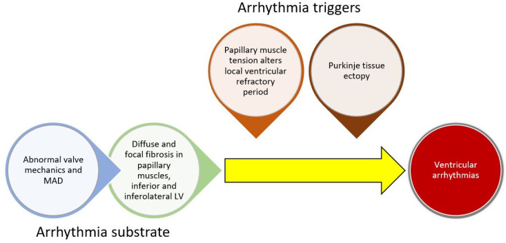
Mitral valve prolapse (MVP) is a common cause of valvular heart disease. Although many patients with MVP have a benign course, there is increasing recognition of an arrhythmic phenotype associated with ventricular arrhythmias and sudden cardiac death (SCD). Pathophysiologic mechanisms associated with arrhythmias include cardiac fibrosis, mechanical stress induced changes in ventricular refractory periods, as well as electrophysiologic changes in Purkinje fibers. Clinically, a variety of risk factors including demographic, electrocardiographic, and imaging characteristics help to identify patients with MVP at the highest at risk of SCD and arrhythmias. Once identified, recent advances in treatment including device therapy, catheter ablation, and surgical interventions show promising outcomes. In this review, we will summarize the incidence of ventricular arrhythmias and SCD in patients with MVP, the association with mitral annular disjunction, mechanisms of arrhythmogenesis, methods for arrhythmic and SCD risk stratification including findings with multimodality imaging, and treatments for the primary and secondary prevention of SCD.

Keywords: catheter ablation; implantable cardiac defibrillator; mitral annular disjunction; mitral valve prolapse; multi-modality imaging; sudden cardiac death.


Conflict of interest statement

The authors declare no conflict of interest.

Figures

Figure 1
Figure 1
Conceptual framework for ventricular arrhythmogenesis involving the development of cardiac substrate and triggers for arrhythmias.
Figure 2
Figure 2
Electrocardiographic findings of arrhythmic mitral valve prolapse. Readings from 12-lead Holter monitoring demonstrating multifocal PVCs as well as T-wave inversions in leads II, III, aVF in a young otherwise healthy woman with bileaflet mitral valve prolapse, mitral annular disjunction, and a high burden of symptomatic premature ventricular contractions. The patient was inducible for sustained polymorphic VT during electrophysiology study and underwent subcutaneous implantable cardiac defibrillator (ICD) implantation.
Figure 3
Figure 3
Echocardiographic findings of arrhythmic mitral valve prolapse and mitral annular disjunction. (A) Transthoracic echocardiography view of bileaflet prolapse seen with mitral annular disjunction (bracket) in the parasternal long axis view. (B) Transesophageal echocardiography view of mitral valve prolapse with color Doppler showing mitral regurgitation in a mid-esophageal long axis view.
Figure 4
Figure 4
Cardiac magnetic resonance findings of arrhythmic mitral valve prolapse and mitral annular disjunction. (A) Bileaflet prolapse seen in a view analogous to apical 4 chamber view. (B) Mitral annular disjunction (bracket) where the mitral valve hinge point separates from the basal left ventricular myocardium and has systolic excursion into the left atrium. (C) Late gadolinium enhancement of a papillary muscle (arrow) seen in a view analogous to parasternal short axis.
Figure 5
Figure 5
Clinical approach to risk stratification for sudden cardiac death or complex ventricular arrhythmias in MVP patients with clinical concern for arrhythmias. Most patients should undergo initial baseline testing with history, transthoracic echocardiography, and resting 12-lead ECG. Cardiac monitoring should be considered in those with a clinical concern for arrhythmias or if the presence of ventricular arrhythmias on monitoring would significantly modify risk categorization. If testing is abnormal or multiple risk factors are seen, cardiac MR can be considered. If there is an abnormal cardiac MR or very high-risk initial history and testing, we utilize a shared decision-making approach to electrophysiology study and possible implantable cardiac defibrillator. Premature ventricular contraction and/or ventricular tachycardia ablation can be considered as well if indicated (see text). TTE: transthoracic echocardiogram; ECG: electrocardiogram; SCD: sudden cardiac death; TWI: T-wave inversions; QTc: corrected QT interval; VT: ventricular tachycardia; PVC: premature ventricular complex; MAD: mitral annular disjunction; MVP: mitral valve prolapse; LV: left ventricle; LA: left atrium; GLS: global longitudinal strain; cardiac MR: cardiac magnetic resonance imaging: LGE: late gadolinium enhancement.
Figure 6
Figure 6
Papillary muscle ablation utilizing cryo-energy and intracardiac echocardiography. (A) Electroanatomic map highlighting cryo-energy lesions to a papillary muscle. (B) Intracardiac echocardiography is a useful adjunct to confirm good contact and stability of catheter to papillary muscle during ablation. PM PAP: posteromedial papillary muscle, LV: left ventricle.

Similar articles

Cited by

References

    1. Criley J.M., Lewis K.B., Humphries J.O., Ross R.S. Prolapse of the mitral valve: Clinical and cine-angiocardiographic findings. Heart. 1966;28:488–496. doi: 10.1136/hrt.28.4.488. - DOI - PMC - PubMed
    1. Freed L.A., Levy D., Levine R.A., Larson M.G., Evans J.C., Fuller D.L., Lehman B., Benjamin E.J. Prevalence and clinical outcome of mitral-valve prolapse. N. Engl. J. Med. 1999;341:1–7. doi: 10.1056/NEJM199907013410101. - DOI - PubMed
    1. Devereux R.B., Jones E.C., Roman M.J., Howard B.V., Fabsitz R.R., Liu J.E., Palmieri V., Welty T.K., Lee E.T. Prevalence and correlates of mitral valve prolapse in a population-based sample of American Indians: The strong heart study. Am. J. Med. 2001;111:679–685. doi: 10.1016/S0002-9343(01)00981-0. - DOI - PubMed
    1. Nalliah C.J., Mahajan R., Elliott A.D., Haqqani H., Lau D.H., Vohra J.K., Morton J.B., Semsarian C., Marwick T., Kalman J.M., et al. Mitral valve prolapse and sudden cardiac death: A systematic review and meta-analysis. Heart. 2019;105:144–151. doi: 10.1136/heartjnl-2017-312932. - DOI - PubMed
    1. Freed L.A., Benjamin E.J., Levy D., Larson M.G., Evans J.C., Fuller D.L., Lehman B., Levine R.A. Mitral valve prolapse in the general population. J. Am. Coll. Cardiol. 2002;40:1298–1304. doi: 10.1016/S0735-1097(02)02161-7. - DOI - PubMed

LinkOut - more resources