. 2022 Feb 17;9(2):62.
doi: 10.3390/jcdd9020062.

DCHS1, Lix1L, and the Septin Cytoskeleton: Molecular and Developmental Etiology of Mitral Valve Prolapse

Affiliations

DCHS1, Lix1L, and the Septin Cytoskeleton: Molecular and Developmental Etiology of Mitral Valve Prolapse

Kelsey S Moore et al. J Cardiovasc Dev Dis. .

Abstract

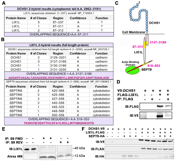
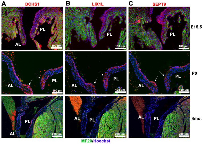
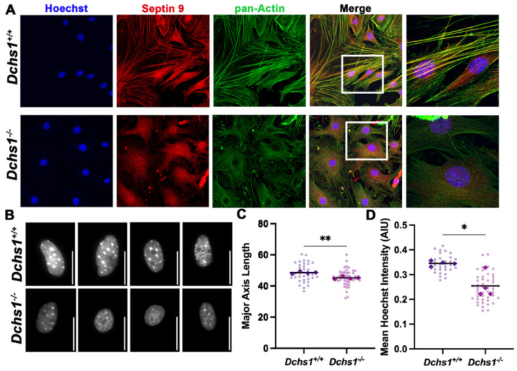
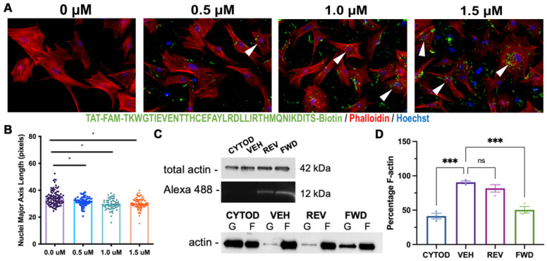
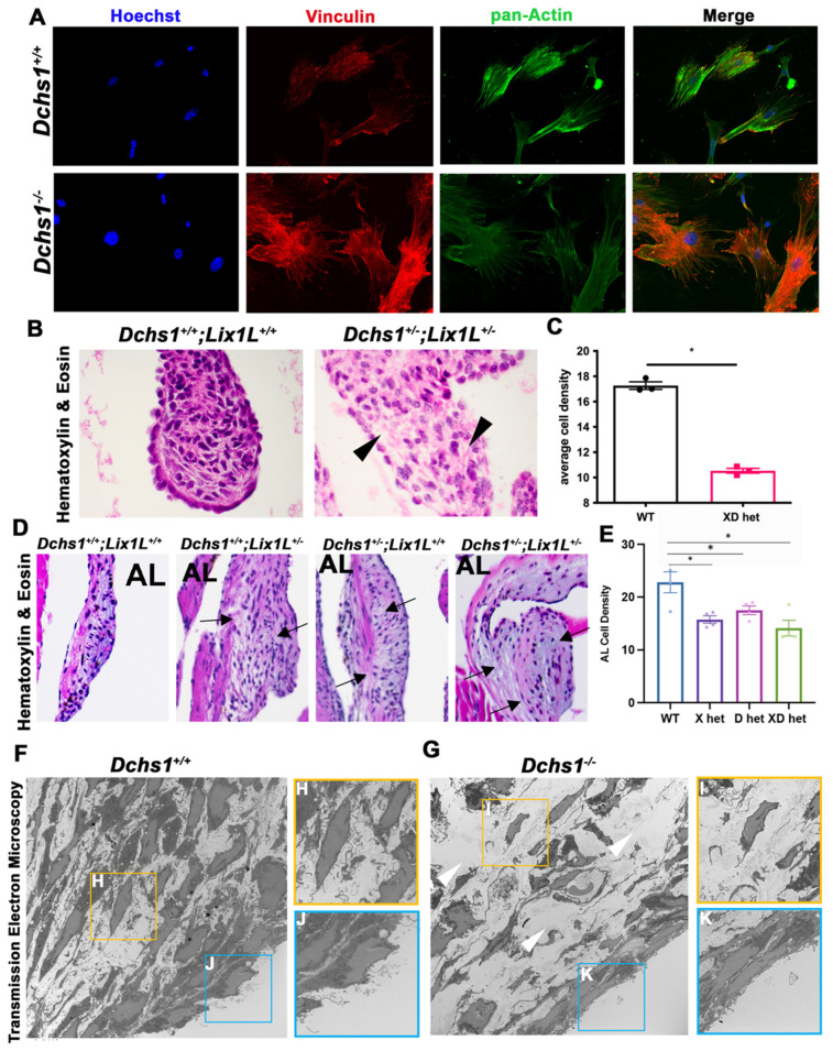
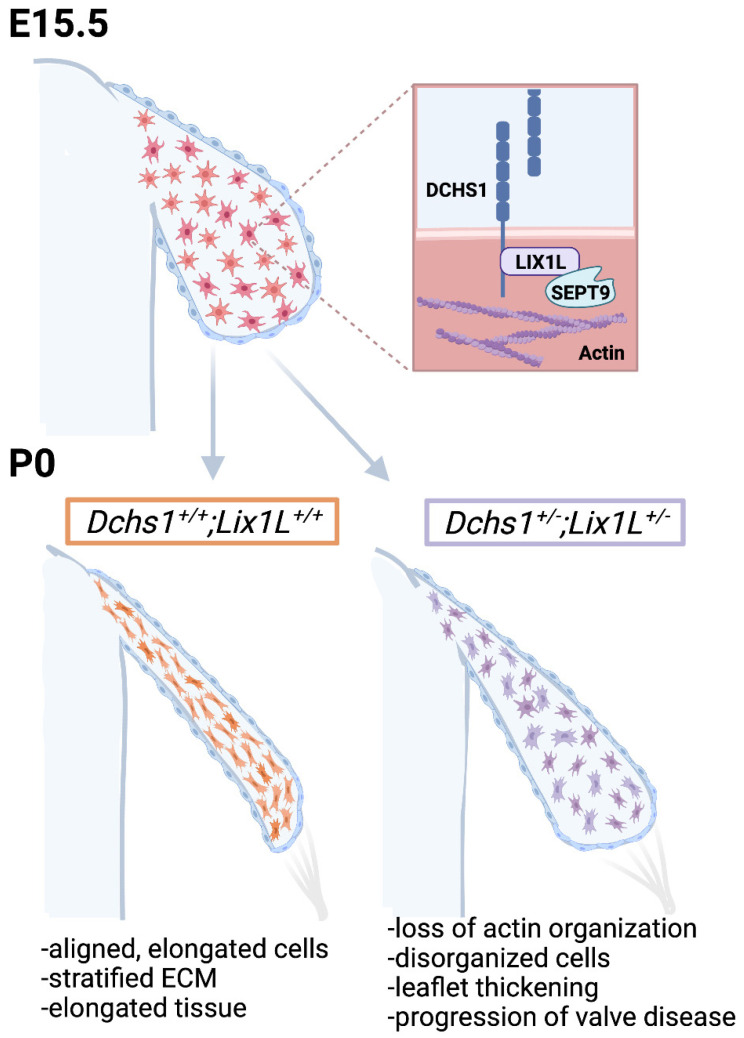
Mitral valve prolapse (MVP) is a common cardiac valve disease that often progresses to serious secondary complications requiring surgery. MVP manifests as extracellular matrix disorganization and biomechanically incompetent tissues in the adult setting. However, MVP has recently been shown to have a developmental basis, as multiple causal genes expressed during embryonic development have been identified. Disease phenotypes have been observed in mouse models with human MVP mutations as early as birth. This study focuses on the developmental function of DCHS1, one of the first genes to be shown as causal in multiple families with non-syndromic MVP. By using various biochemical techniques as well as mouse and cell culture models, we demonstrate a unique link between DCHS1-based cell adhesions and the septin-actin cytoskeleton through interactions with cytoplasmic protein Lix1-Like (LIX1L). This DCHS1-LIX1L-SEPT9 axis interacts with and promotes filamentous actin organization to direct cell-ECM alignment and valve tissue shape.

Keywords: DCHS1; cytoskeleton; heart valve development; mitral valve prolapse; septin.


Conflict of interest statement

The authors declare no conflict of interest.

Figures

Figure 1
Figure 1
Identification of DCHS1-LIX1L-SEPT9 (DLS) protein complex. (A) Yeast two-hybrid (Y2H) screen using cytoplasmic tail (A.A. 2962–3191) of Dachsous Cadherin Related-1 (DCHS1) as bait reveals Lix1-Like (LIX1L). (B) Y2H with full-length LIX1-Like (LIX1L) identifies DCHS1 and septin-9 (SEPT9) as binding partners. In A and B, confidence scores represent likelihood of an interaction, with A being the highest level of confidence (See Methods for details). (C) Diagram depicting interacting proteins and binding domains. (D) Co-Immunoprecipitation (co-IP) and immunoblotting (IB) analysis with DCHS1-V5 and LIX1L-FLAG transfected in HEK293T cells confirms protein interaction. (E) Co-IP and IB of cVIC protein lysate treated with 10 μg peptide mimicking the LIX1L-SEPT9 binding domain (S9-FWD:5-FAM-YGRKKRRQRRR-Ahx TKWGTIEVENTTHCEFAYLRDLLIRTHMQNIKDIT-Lys(Biotin), S9-REV:5-FAM-YGRKKRRQRRR-Ahx-TIDKINQMHTRILLDRLYAFECHTTNEVEITGWKT-Lys(Biotin)). (F) IB of DCHS1-V5, LIX1L-FLAG and SEPT9-HA co-transfected into HEK293T cells depicts stabilization of DCHS1 protein only in the presence of LIX1L.
Figure 2
Figure 2
Spatio-temporal expression of DCHS1-LIX1L-SEPT9 proteins throughout development. IHC staining of (A) DCHS1, (B) LIX1L and (C) SEPT9 (red), MF20 (green), and nuclei (Hoechst, blue) on sister sections of E15.5, P0 and four-month-old wildtype mouse valve tissue reveals similar expression throughout the interstitium with localization to the leaflet tips by P0 (arrows) and restriction to the endothelium through adulthood. AL = anterior leaflet, PL = posterior leaflet.
Figure 3
Figure 3
Epistasis analysis reveals DCHS1-LIX1L genetic interaction at postnatal day zero. (AD) Hematoxylin and eosin (H&E) staining and (EH) 3D reconstructions of postnatal day zero (P0) mitral valves isolated from wildtype (W), single heterozygote (LIX1L+/−, L or DCHS1+/−, D) and compound heterozygote (LIX1L+/−;DCHS1+/−, LD) mice reveal thickening of single heterozygote and compound heterozygote anterior leaflets (AL) compared to wildtype control littermates. (IK) Total mitral valve volume measurements from 3D-reconstructed leaflet surfaces demonstrate a significant increase in compound heterozygotes AL leaflets compared to wildtype, with no significance observed in posterior leaflet (PL) volumes. Two-dimensional measurements of length (L,N) and width (M,O) along the leaflet reveals that changes in leaflet volume are due to significant leaflet thickening in the base and tip of the (L,M) anterior leaflets of compound heterozygotes versus single heterozygotes or wildtypes. (N,O) No significant differences were observed in the posterior leaflet of these mice. N = 4–5 animals per genotype, 2D measurements completed on four sections per animal and depicted by dots, averages are plotted as diamonds, * p < 0.05, ** p < 0.005 with One-Way Anova.
Figure 4
Figure 4
Adult heterozygotes display mitral valve disease phenotype of myxomatous degeneration. (A) Representative H&E staining of 11-month-old wildtype (W), single (L, D) and compound heterozygote (LD) mitral valves depict thickening and bulging of single and compound heterozygotes anterior and posterior leaflets (AL, PL) (black arrows). Immunohistochemical (IHC) staining of (B) Versican (red) and (C) Collagen 1α1 (red), myocardium (MF20, green) and nuclei (Hoechst, blue) reveals increased extracellular matrix deposition and disorganization (white arrows and arrowheads) in single or compound heterozygotes. (D,E) Quantification of ECM staining intensity reveals statistically significant increases in single or compound heterozygote leaflets compared to wildtype controls, indications of myxomatous degeneration. N = 3 per genotype, depicted with mean and SEM, * p < 0.05, ** p < 0.005 with One Way Anova.
Figure 5
Figure 5
Septin-actin defects in DCHS1-deficient fibroblasts. (A) Wildtype (DCHS1+/+) and knock-out, (DCHS1−/−), cardiac fibroblasts (CFs) were seeded on collagen coated slides and stained for pan-actin (green), septin 9 (red) and nuclei (blue). In DCHS1−/− fibroblasts, actin and SEPT9 no longer co-localize coincident with loss in stress fiber organization and altered cell shape. (B) Representative grayscale images of nuclei stained with Hoechst illustrate a rounded phenotype in DCHS-−/−, scale bar = 50 pixels. (C) Quantification of nuclei major axis length (pixels) and (D) Hoechst staining intensity (Arbitrary intensity units, AIU) reveals significant reductions in knockout cells compared to wildtype controls suggesting loss of intracellular tension. 30–50 cells imaged per genotype, N = 4 ** p < 0.005, * p < 0.05 with Student’s t-test.
Figure 6
Figure 6
Targeting the LIX1L-SEPT9 binding domain with decoy peptide disrupts F-actin polymerization. (A) cVICs treated with increasing doses of SEPT9 decoy peptide conjugated with cell permeant TAT sequence= GRKKRRQRRRPQ, 5-FAM and biotin, diluted in DMSO (0–1.5 μM) for 2 hrs and seeded on collagen coated slides prior to immunocytochemistry staining with phallodin-594 (red) and Hoechst (nuclei = blue). Actin bundling is observed in cells containing peptide (arrowheads). (B) Quantification of nuclei major axis length reveals significant reductions in nuclei elongation in response to peptide treatment. N = 3 per treatment group with 20–30 cells per treatment, * p < 0.05 with One-Way Anova. (C) F/G-actin polymerization assay performed on cVICs seeded at 50% confluence, treated with cytochalasin-D (CYTOD, 1 μM) for 1.5 h and rescued with serum containing media with vehicle (DMSO, VEH), forward (FWD) or reverse (REV) peptide at 1.5 μM. IB of representative total actin and peptide loading and separated F/G-actin content. (D) Quantification of F-actin percentage illustrates reduction in F-actin polymerization in FWD peptide-treated cells compared to vehicle. n = 3 per treatment, *** p < 0.0005 with Ordinary One-Way Anova.
Figure 7
Figure 7
Consequences of actin defects. (A) ICC of vinculin (red), pan-actin (green), and nuclei (Hoechst, blue) in wildtype (DCHS1+/+) and DCHS1 KO (DCHS1−/−) reveals loss of stress fiber formation and vinculin organization and localization to focal adhesions. (B) H&E images of wildtype and compound heterozygote P0 valves depict increased density of ECM between interstitial cells (black arrowheads), also observed in (D) 11-month-old leaflets of single and compound heterozygote anterior leaflets (arrows). (C,E) Quantification of cell density measured by cells divided by total leaflet area reveals significant decreases in compound heterozygotes (XD het) compared to controls at P0 and both single and compound heterozygotes in adults. Graphs depict average cell density per animal with error bars of SEM, n = 3–4 animals per genotype, * p < 0.005. (F,G) Transmission electron microscopy (TEM) images of wildtype and DCHS1 KO anterior leaflets at P0 show increased ECM content in DCHS1 KOs and less dense interstitium with round nuclei and loss of filopodia-structures (yellow boxes, (H,I)). Filopodia-like structures appear to be lost along the DCHS1 KO endothelium (blue boxes, J,K).
Figure 8
Figure 8
Working model of valve morphogenesis. DCHS1, LIX1L and Sept9 are expressed in undifferentiated mesenchymal cells during embryonic development that are randomly distributed throughout the tissue. As these cells proliferate and increase in density, they encounter neighboring cells and engage cadherin receptors, much like train cars hooking together on a railroad track (ECM). DCHS1-LIX1L-SEPT9 (DLS) complex promotes filamentous actin formation and cell elongation. All cargo attached to the cell membrane, such as integrins and the ECM, then passively assume a similar organization and shape as the cells. This results in aligned, elongated cells and tissues with a stratified ECM. Defects in any step of this process would be anticipated as resulting in disrupted actin organization, disorganized cells, leaflet thickening and progression to a valve pathology.

Similar articles

Cited by

References

    1. Gammie J.S., Chikwe J., Badhwar V., Thibault D.P., Vemulapalli S., Thourani V.H., Gillinov M., Adams D.H., Rankin J.S., Ghoreishi M., et al. Isolated Mitral Valve Surgery: The Society of Thoracic Surgeons Adult Cardiac Surgery Database Analysis. Ann. Thorac. Surg. 2018;106:716–727. doi: 10.1016/j.athoracsur.2018.03.086. - DOI - PubMed
    1. Grigioni F., Tribouilloy C., Avierinos J.F., Barbieri A., Ferlito M., Trojette F., Tafanelli L., Branzi A., Szymanski C., Habib G., et al. Outcomes in Mitral Regurgitation Due to Flail Leaflets: A Multicenter European Study. JACC Cardiovasc. Imaging. 2008;1:133–141. doi: 10.1016/j.jcmg.2007.12.005. - DOI - PubMed
    1. Durst R., Sauls K., Peal D.S., DeVlaming A., Toomer K., Leyne M., Salani M., Talkowski M., Brand H., Perrocheau M., et al. Mutations in DCHS1 cause mitral valve prolapse. Nature. 2015;525:109–113. doi: 10.1038/nature14670. - DOI - PMC - PubMed
    1. Clemenceau A., Bérubé J.-C., Bélanger P., Gaudreault N., Lamontagne M., Toubal O., Clavel M.-A., Capoulade R., Mathieu P., Pibarot P., et al. Deleterious variants in DCHS1 are prevalent in sporadic cases of mitral valve prolapse. Mol. Genet. Genom. Med. 2017;6:114–120. doi: 10.1002/mgg3.347. - DOI - PMC - PubMed
    1. Rogulja D., Rauskolb C., Irvine K.D. Morphogen Control of Wing Growth through the Fat Signaling Pathway. Dev. Cell. 2008;15:309–321. doi: 10.1016/j.devcel.2008.06.003. - DOI - PMC - PubMed

LinkOut - more resources