

Leukocyte-Mediated Cardiac Repair after Myocardial Infarction in Non-Regenerative vs. Regenerative Systems
- PMID: 35200716
- PMCID: PMC8877434
- DOI: 10.3390/jcdd9020063
Leukocyte-Mediated Cardiac Repair after Myocardial Infarction in Non-Regenerative vs. Regenerative Systems
Abstract
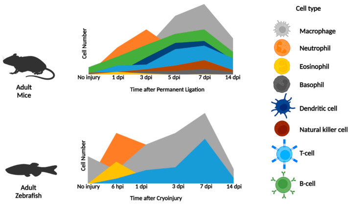
Innate and adaptive leukocytes rapidly mobilize to ischemic tissues after myocardial infarction in response to damage signals released from necrotic cells. Leukocytes play important roles in cardiac repair and regeneration such as inflammation initiation and resolution; the removal of dead cells and debris; the deposition of the extracellular matrix and granulation tissue; supporting angiogenesis and cardiomyocyte proliferation; and fibrotic scar generation and resolution. By organizing and comparing the present knowledge of leukocyte recruitment and function after cardiac injury in non-regenerative to regenerative systems, we propose that the leukocyte response to cardiac injury differs in non-regenerative adult mammals such as humans and mice in comparison to cardiac regenerative models such as neonatal mice and adult zebrafish. Specifically, extensive neutrophil, macrophage, and T-cell persistence contributes to a lengthy inflammatory period in non-regenerative systems for adverse cardiac remodeling and heart failure development, whereas their quick removal supports inflammation resolution in regenerative systems for new contractile tissue formation and coronary revascularization. Surprisingly, other leukocytes have not been examined in regenerative model systems. With this review, we aim to encourage the development of improved immune cell markers and tools in cardiac regenerative models for the identification of new immune targets in non-regenerative systems to develop new therapies.
Keywords: cardiac injury; heart; humans; immune cells; inflammatory; leukocyte; mice; myocardial infarction; regeneration; repair; zebrafish.
Conflict of interest statement
The authors declare no conflict of interest.
Figures

Similar articles
-
Comparative single-cell profiling reveals distinct cardiac resident macrophages essential for zebrafish heart regeneration.Elife. 2023 Jul 27;12:e84679. doi: 10.7554/eLife.84679. Elife. 2023. PMID: 37498060 Free PMC article.
-
The immune system and cardiac repair.Pharmacol Res. 2008 Aug;58(2):88-111. doi: 10.1016/j.phrs.2008.06.007. Epub 2008 Jun 24. Pharmacol Res. 2008. PMID: 18620057 Free PMC article. Review.
-
Decellularized neonatal cardiac extracellular matrix prevents widespread ventricular remodeling in adult mammals after myocardial infarction.Acta Biomater. 2019 Mar 15;87:140-151. doi: 10.1016/j.actbio.2019.01.062. Epub 2019 Jan 30. Acta Biomater. 2019. PMID: 30710713 Free PMC article.
-
The Biological Basis for Cardiac Repair After Myocardial Infarction: From Inflammation to Fibrosis.Circ Res. 2016 Jun 24;119(1):91-112. doi: 10.1161/CIRCRESAHA.116.303577. Circ Res. 2016. PMID: 27340270 Free PMC article. Review.
-
Emerging Roles for Immune Cells and MicroRNAs in Modulating the Response to Cardiac Injury.J Cardiovasc Dev Dis. 2019 Jan 15;6(1):5. doi: 10.3390/jcdd6010005. J Cardiovasc Dev Dis. 2019. PMID: 30650599 Free PMC article. Review.
Cited by
-
Cross-species single-cell RNA-seq analysis reveals disparate and conserved cardiac and extracardiac inflammatory responses upon heart injury.Commun Biol. 2024 Dec 3;7(1):1611. doi: 10.1038/s42003-024-07315-x. Commun Biol. 2024. PMID: 39627536 Free PMC article.
-
Quo Vadis? Immunodynamics of Myeloid Cells after Myocardial Infarction.Int J Mol Sci. 2022 Dec 13;23(24):15814. doi: 10.3390/ijms232415814. Int J Mol Sci. 2022. PMID: 36555456 Free PMC article. Review.
-
Comparative Analysis of Heart Regeneration: Searching for the Key to Heal the Heart-Part II: Molecular Mechanisms of Cardiac Regeneration.J Cardiovasc Dev Dis. 2023 Aug 22;10(9):357. doi: 10.3390/jcdd10090357. J Cardiovasc Dev Dis. 2023. PMID: 37754786 Free PMC article. Review.
-
Mesenchymal stem cells derived exosomes: a new era in cardiac regeneration.Stem Cell Res Ther. 2025 Jan 23;16(1):16. doi: 10.1186/s13287-024-04123-2. Stem Cell Res Ther. 2025. PMID: 39849585 Free PMC article. Review.
-
PPAR Alpha Activation by Clofibrate Alleviates Ischemia/Reperfusion Injury in Metabolic Syndrome Rats by Decreasing Cardiac Inflammation and Remodeling and by Regulating the Atrial Natriuretic Peptide Compensatory Response.Int J Mol Sci. 2023 Mar 10;24(6):5321. doi: 10.3390/ijms24065321. Int J Mol Sci. 2023. PMID: 36982395 Free PMC article.
References
-
- Centers for Disease Control and Prevention . Underlying Cause of Death, 1999–2018. Centers for Disease Control and Prevention; Atlanta, GA, USA: 2018. [(accessed on 12 March 2020)]. CDC WONDER Online Database. Available online: https://wonder.cdc.gov/ucd-icd10.html.
-
- Virani S.S., Alonso A., Aparicio H.J., Benjamin E.J., Bittencourt M.S., Callaway C.W., Carson A.P., Chamberlain A.M., Cheng S., Delling F.N., et al. Heart Disease and Stroke Statistics-2021 Update: A Report from the American Heart Association. Circulation. 2021;143:e254–e743. doi: 10.1161/CIR.0000000000000950. - DOI - PubMed
Publication types
Grants and funding
LinkOut - more resources
Full Text Sources
Other Literature Sources
