

Monte Carlo Characterization of the Trimage Brain PET System
- PMID: 35200724
- PMCID: PMC8878795
- DOI: 10.3390/jimaging8020021
Monte Carlo Characterization of the Trimage Brain PET System
Abstract
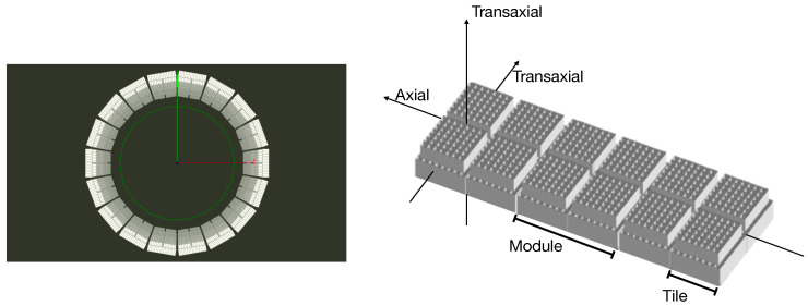
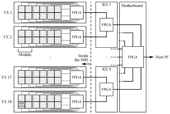
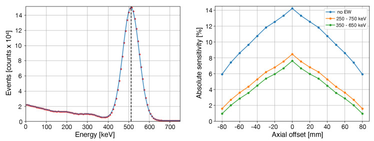
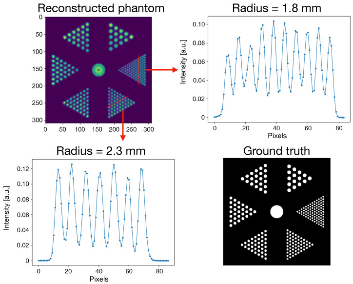
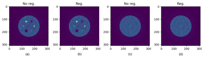
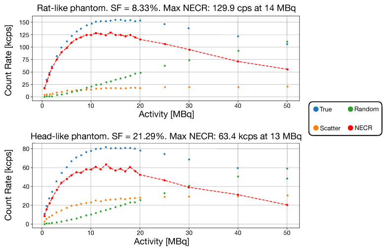
The TRIMAGE project aims to develop a brain-dedicated PET/MR/EEG (Positron Emission Tomography/Magnetic Resonance/Electroencephalogram) system that is able to perform simultaneous PET, MR and EEG acquisitions. The PET component consists of a full ring with 18 sectors. Each sector includes three square detector modules based on dual sstaggered LYSO:Ce matrices read out by SiPMs. Using Monte Carlo simulations and following NEMA (National Electrical Manufacturers Association) guidelines, image quality procedures have been applied to evaluate the performance of the PET component of the system. The performance are reported in terms of spatial resolution, uniformity, recovery coefficient, spill over ratio, noise equivalent count rate (NECR) and scatter fraction. The results show that the TRIMAGE system is at the top of the current brain PET technologies.
Keywords: Monte Carlo characterization; NEMA; PET/MR; TRIMAGE project; brain PET.
Conflict of interest statement
The authors declare no conflict of interest.
Figures
Similar articles
-
Monte Carlo evaluation of hypothetical long axial field-of-view PET scanner using GE Discovery MI PET front-end architecture.Med Phys. 2022 Feb;49(2):1139-1152. doi: 10.1002/mp.15422. Epub 2022 Jan 10. Med Phys. 2022. PMID: 34954831
-
Monte Carlo Simulations of the GE Signa PET/MR for Different Radioisotopes.Front Physiol. 2020 Sep 15;11:525575. doi: 10.3389/fphys.2020.525575. eCollection 2020. Front Physiol. 2020. PMID: 33041852 Free PMC article.
-
GATE Monte Carlo simulations for variations of an integrated PET/MR hybrid imaging system based on the Biograph mMR model.Phys Med Biol. 2015 Jun 21;60(12):4731-52. doi: 10.1088/0031-9155/60/12/4731. Epub 2015 Jun 4. Phys Med Biol. 2015. PMID: 26040657
-
Performance characteristic evaluation of a bismuth germanate-based high-sensitivity 5-ring discovery image quality positron emission tomography/computed tomography system as per National Electrical Manufacturers Association NU 2-2012.World J Nucl Med. 2019 Dec 18;18(4):351-360. doi: 10.4103/wjnm.WJNM_72_18. eCollection 2019 Oct-Dec. World J Nucl Med. 2019. PMID: 31933550 Free PMC article.
-
Dosage optimization in positron emission tomography: state-of-the-art methods and future prospects.Am J Nucl Med Mol Imaging. 2015 Oct 12;5(5):527-47. eCollection 2015. Am J Nucl Med Mol Imaging. 2015. PMID: 26550543 Free PMC article. Review.
Cited by
-
The quest for multifunctional and dedicated PET instrumentation with irregular geometries.Ann Nucl Med. 2024 Jan;38(1):31-70. doi: 10.1007/s12149-023-01881-6. Epub 2023 Nov 12. Ann Nucl Med. 2024. PMID: 37952197 Free PMC article. Review.
References
LinkOut - more resources
Full Text Sources
