Review
. 2022 Feb 1;8(2):35.
doi: 10.3390/jimaging8020035.

Multimodality Imaging in Ischemic Chronic Cardiomyopathy

Affiliations
Review

Multimodality Imaging in Ischemic Chronic Cardiomyopathy

Giuseppe Muscogiuri et al. J Imaging. .

Abstract

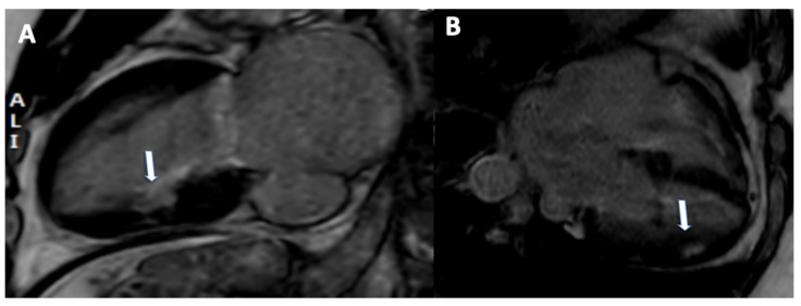
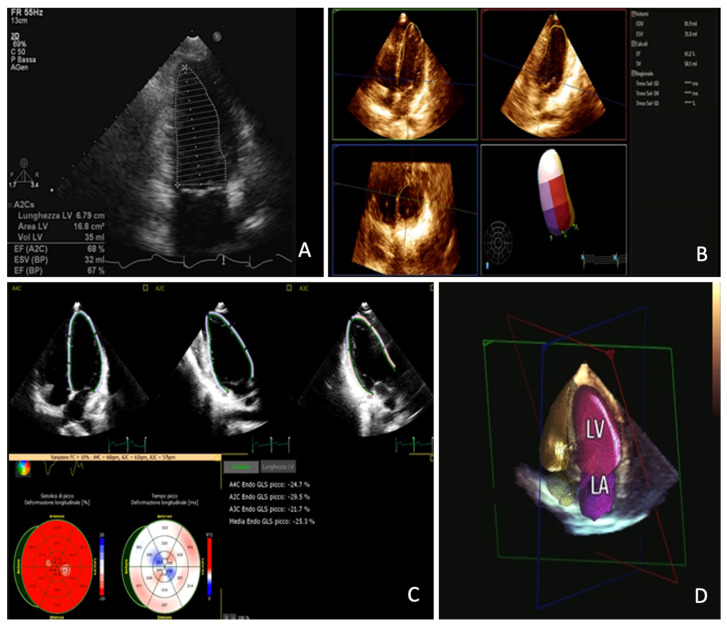
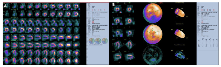
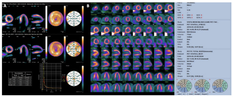
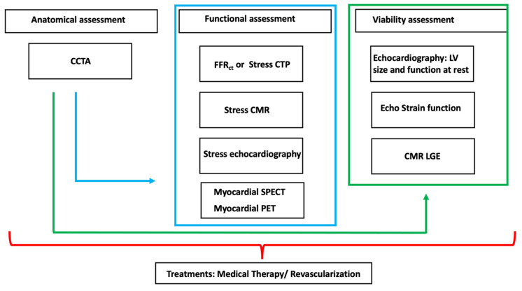
Ischemic chronic cardiomyopathy (ICC) is still one of the most common cardiac diseases leading to the development of myocardial ischemia, infarction, or heart failure. The application of several imaging modalities can provide information regarding coronary anatomy, coronary artery disease, myocardial ischemia and tissue characterization. In particular, coronary computed tomography angiography (CCTA) can provide information regarding coronary plaque stenosis, its composition, and the possible evaluation of myocardial ischemia using fractional flow reserve CT or CT perfusion. Cardiac magnetic resonance (CMR) can be used to evaluate cardiac function as well as the presence of ischemia. In addition, CMR can be used to characterize the myocardial tissue of hibernated or infarcted myocardium. Echocardiography is the most widely used technique to achieve information regarding function and myocardial wall motion abnormalities during myocardial ischemia. Nuclear medicine can be used to evaluate perfusion in both qualitative and quantitative assessment. In this review we aim to provide an overview regarding the different noninvasive imaging techniques for the evaluation of ICC, providing information ranging from the anatomical assessment of coronary artery arteries to the assessment of ischemic myocardium and myocardial infarction. In particular this review is going to show the different noninvasive approaches based on the specific clinical history of patients with ICC.

Keywords: cardiac magnetic resonance; chronic ischemic cardiomyopathy; computed tomography angiography; echocardiography; multimodality imaging; nuclear medicine.


Conflict of interest statement

The authors declare no conflict of interest.

Figures

Figure 1
Figure 1
A 77-year-old male with in-stent restenosis in the proximal segment of the left anterior descending artery (A), a filling defect in the proximal part of the stent (arrow) is clearly visualized. The invasive coronary angiogram (B) shows in-stent restenosis in the stent located in the proximal left anterior descending artery (arrow).
Figure 2
Figure 2
A 70-year-old female patient underwent to CCTA for chest pain. Right coronary artery (A) and left circumflex artery (D) were free from significant stenosis; whereas left anterior descending artery shows moderate proximal stenosis ((B), arrow). Ramus intermedius shows a severe, fibro-lipid plaque stenosis ((C), arrowhead). The FFRCT assessment confirmed the functional significance of the stenosis proximal-ramus intermedius ((E), arrowhead), whereas the FFRCT values of the left anterior descending artery were above the ischemia threshold of 0.80. The invasive coronary angiogram ((F), arrowhead) shows severe stenosis of the proximal tract of the ramus intermedius.
Figure 3
Figure 3
A 60-year-old female patient underwent to CCTA and dynamic CT perfusion for suspected coronary artery disease. Diffuse coronary calcification on left anterior descending artery (A), left circumflex (B) and right coronary (C), showing mild reduction of flow in dynamic CT perfusion on lateral wall, in particular at the apical segment (D).
Figure 4
Figure 4
A 65-year-old male patient with known history of chronic total occlusion of right coronary artery, moderate stenosis on left anterior descending artery and mild stenosis on right coronary artery. Rest perfusion sequence does not show any significant defect of perfusion (A). During stress acquisition, a deficit of perfusion was observed in the septum and inferior wall ((B), arrows).
Figure 5
Figure 5
A 45-year-old patient with history of myocardial infarction. Subendocardial late gadolinium enhancement was observed in inferior (arrow, (A)) and inferolateral (arrow, (B)).
Figure 6
Figure 6
Panel (A): bi-dimensional echocardiography measurement of left ventricle ejection fraction; Panel (B): three-dimensional echocardiography measurement of left ventricle ejection fraction; Panel (C): Left Ventricle global longitudinal strain assessment; Panel (D): three-dimensional left ventricle and left atrium automatic measurement with artificial intelligence algorithm.
Figure 7
Figure 7
Myocardial perfusion SPECT with Tc-99m-Tetrofosmin in a patient with CAD performed at stress (maximal cycloergometer exercise) and at rest. Stress and rest slices in three axes are shown in (Panel (A)). Polar maps under stress and at rest are shown in (Panel (B)). Slices and polar maps show a stress perfusion defect in the inferior wall which significantly improves (reverses) in the rest study. The pattern is typical of stress-induced myocardial ischemia.
Figure 8
Figure 8
(Panel (A)) dynamic myocardial 13N-ammonia PET/CT in a normal patient. First row shows representative left ventricular perfusion myocardial slices under maximal vasodilator stress (with quantitative myocardial stress flow values); second row shows representative left ventricular perfusion myocardial slices at rest (with quantitative myocardial rest flow values). Absolute flow values are presented in the table and flow reserve is automatically calculated for the different territories and also for the whole left ventricle (global). (Panel (B)) myocardial 13N-ammonia PET/CT stress/rest perfusion study in the same patient. The distribution of perfusion is homogeneous both at stress and at rest.
Figure 9
Figure 9
Multimodality evaluation of patients with ICC. Green arrows shows the sequential evaluation of coronary anatomy followed by the assessment of myocardial viability. Blue arrow shows sequential approach for the anatomical evaluation followed by functional assessment. Each approach should be customized considering the local expertise and the clinical needs.

Similar articles

Cited by

References

    1. Knuuti J., Wijns W., Saraste A., Capodanno D., Barbato E., Funck-Brentano C., Prescott E., Storey R.F., Deaton C., Cuisset T., et al. 2019 ESC Guidelines for the diagnosis and management of chronic coronary syndromes. Eur. Heart J. 2020;41:407–477. doi: 10.1093/eurheartj/ehz425. - DOI - PubMed
    1. Muscogiuri G., Ricci F., Scafuri S., Guglielmo M., Baggiano A., De Stasio V., Di Donna C., Spiritigliozzi L., Chiocchi M., Lee S.J., et al. Cardiac Magnetic Resonance Tissue Characterization in Ischemic Cardiomyopathy. J. Thorac. Imaging. 2021;37:2–16. doi: 10.1097/RTI.0000000000000621. - DOI - PubMed
    1. Andreini D., Conte E., Serruys P.W. Coronary plaque features on CTA can identify patients at increased risk of cardiovascular events. Curr. Opin. Cardiol. 2021;36:784–792. doi: 10.1097/HCO.0000000000000917. - DOI - PubMed
    1. Kadoglou N.P.E., Papadopoulos C.H., Papadopoulos K.G., Karagiannis S., Karabinos I., Loizos S., Theodosis-Georgilas A., Aggeli K., Keramida K., Klettas D., et al. Updated knowledge and practical implementations of stress echocardiography in ischemic and non-ischemic cardiac diseases: An expert consensus of the Working Group of Echocardiography of the Hellenic Society of Cardiology. Hellenic J. Cardiol. 2021 doi: 10.1016/j.hjc.2021.07.006. In Press . - DOI - PubMed
    1. Feldman D.I., Latina J., Lovell J., Blumenthal R.S., Arbab-Zadeh A. Coronary computed tomography angiography in patients with stable coronary artery disease. Trends Cardiovasc. Med. 2021 doi: 10.1016/j.tcm.2021.08.009. - DOI - PubMed