Review
. 2022 May 24;34(6):2120-2139.
doi: 10.1093/plcell/koac069.

Division site determination during asymmetric cell division in plants

Affiliations
Review

Division site determination during asymmetric cell division in plants

Peishan Yi et al. Plant Cell. .

Abstract

During development, both animals and plants exploit asymmetric cell division (ACD) to increase tissue complexity, a process that usually generates cells dissimilar in size, morphology, and fate. Plants lack the key regulators that control ACD in animals. Instead, plants have evolved two unique cytoskeletal structures to tackle this problem: the preprophase band (PPB) and phragmoplast. The assembly of the PPB and phragmoplast and their contributions to division plane orientation have been extensively studied. However, how the division plane is positioned off the cell center during asymmetric division is poorly understood. Over the past 20 years, emerging evidence points to a critical role for polarly localized membrane proteins in this process. Although many of these proteins are species- or cell type specific, and the molecular mechanism underlying division asymmetry is not fully understood, common features such as morphological changes in cells, cytoskeletal dynamics, and nuclear positioning have been observed. In this review, we provide updates on polarity establishment and nuclear positioning during ACD in plants. Together with previous findings about symmetrically dividing cells and the emerging roles of developmental cues, we aim to offer evolutionary insight into a common framework for asymmetric division-site determination and highlight directions for future work.


Figures

Figure 1
Figure 1
The default mechanism of division site determination during symmetric cell division. In most plant cells, the nucleus occupies a large cytoplasmic area at the cell center. In vacuolate cells, the nucleus is off-center and has to migrate to the cell center before mitosis. This process is mediated by MTs (gray lines) that connect the nuclear membrane and the cell cortex (1). At preprophase, a ring-shaped PPB (blue circle) is assembled by the realignment of cortical MTs (2). At prometaphase, the PPB is disassembled (3). A series of MT-associated proteins that have been recruited to the cortical ring following PPB assembly are maintained. These proteins collectively mark the CDZ (orange circle) (3). At anaphase, with the aid of the mitotic spindle, the assembly of the cell plate (magenta disk) is initiated at the cell center. Later on, the mitotic spindle is transformed into a phragmoplast. The assembling cell plate expands toward the CDZ under the guidance of the phragmoplast (4). Eventually, the cell plate fuses with the parental cell membrane at the place labeled by the CDZ (5).
Figure 2
Figure 2
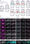
Division site determination during ACD. A, In many plant cells, cell polarization is a prerequisite for initiating asymmetric division (1). Once polarized, the cell undergoes polar cell expansion and migrates its nucleus to an off-center position (2). This process relies on polarity proteins (purple) and cytoskeletal elements (gray lines). However, the responsible molecules can be different depending on the cell type. In the majority of cells in flowering plants, the PPB (blue circle) is subsequently assembled around the nucleus and facilitates the establishment of the CDZ (orange circle), which also occurs during symmetric divisions (3, upper). The CDZ then guides phragmoplast-mediated cell plate (magenta disk) expansion (4–6, upper). In some types of cells such as moss protonema cells and gametophore initials, the PPB is not formed. Instead, the CDZ itself is established after nuclear envelope breakdown (3, lower). In addition, both the mitotic spindle and the CDZ can rotate, thus generating an oriented division plane (4–6, lower). Note that CDZ assembly occurs earlier in Arabidopsis than in mosses and that the rotation of the CDZ has only been reported in moss caulonema tip cells, whose functional significance has not been addressed (see Figure 2B). B, Dynamic establishment of the CDZ in P. patens caulonema tip cells. Time-lapse images show the localization of PpKin12-Ie (a homolog of POKs, left) and PpREN (the homolog of PHGAPs, right). Magenta arrowheads indicate the position of the CDZ. The CDZ localization of PpKin12-Ie and PpREN at metaphase or meta/anaphase (yellow arrowheads) is overlaid with their localization at telophase (red arrowheads) for comparison. Dashed lines show the orientation of the CDZ. Time is shown following nuclear envelope breakdown (0 min). Images are reproduced from Movie S6 in Miki et al. (2014) and Video S5 in Yi and Goshima (2020). Scale bars: 10 µm.
Figure 3
Figure 3
Cell models for studying ACD in Arabidopsis. A, Zygotic division. After fertilization, the zygote exhibits rapid polar growth. Concurrently, its nucleus migrates toward the apical cytoplasm (green arrow) and an expanding vacuole occupies the basal cytoplasm. The PPB is then assembled around the nucleus (blue circle). As the position of the nucleus is asymmetric, the subsequent division produces a small apical cell and a large basal cell, which will develop into the proembryo and suspensor, respectively. B, Founder cell division. Lateral roots are initiated by the division of pairs of founder cells. Before division, two abutting founder cells expand radially. The expansion near the common wall (central domain) is faster than that at the periphery domain. During cell expansion, their nuclei migrate toward the common wall (green arrows) and divide asymmetrically to produce two central cells and two peripheral cells that differ significantly in shape. C, The division of MMCs. The premitotic MMC is polarized with the BASL-associated protein complex on the membrane (purple). This polarity crescent instructs the opposing movement of the nucleus (green arrow). The PPB is formed around the nucleus (blue line) and marks the asymmetric division site. The polarity crescent is inherited by the large daughter cell. After division, the nucleus in the large daughter cell migrates toward the polarity axis. This process is also controlled by the polarity signal.
Figure 4
Figure 4
The division of stomatal SMCs in maize. Before division, the maize stomatal SMCs are polarized by signals from the closely associated GMCs. The SMCs undergo local cell expansion and form a polarized domain at the GMC contact site. The polarization process depends on polarity proteins (PAN1/2 and ROP2/9) and an actin patch (purple). Subsequently, their nuclei migrate toward the polarized cortex (green arrow). As the protrusion of the SMCs is limited in space, the PPB is assembled in a curved shape (blue line). After nuclear envelope breakdown, the mitotic spindle is anchored to the polarized cortex. Cytokinesis proceeds under the guidance of the phragmoplast and generates a lens-shaped small subsidiary cell.
Figure 5
Figure 5
Cell models for studying ACD in P. patens. A, The division of caulonema tip cells. The tip cell exhibits polarized growth under the control of the polarity proteins ROP GTPases (purple). During growth, the tip cell nucleus moves to the cell center (green arrow) and then undergoes mitosis when the cell length reaches approximately twice the length of subapical cells. The PPB is not formed. Instead, the mitotic spindle and CDZ (orange) can rotate to generate an oblique division plane. Although the resulting daughter cells are equal in size, they show remarkable differences in cellular contents and cell fates. B, The division of subapical cells. Subapical cells first initiate a polarized bulge at the apical end (purple), a process that depends on ROP GTPases. During bulging, the nucleus undergoes directed migration (green arrow) and is eventually located in the bulge. Subsequently, the cell divides asymmetrically to produce a small side-branch initial cell. After division, the nucleus in the large daughter cell moves back to the cell center (green arrow). The PPB is absent during subapical cell division, but the CDZ (orange) is retained. C, Localization of the CDZ component PpREN (the moss homolog of PHGAPs) in a subapical cell (yellow arrows) during cytokinesis. Scale bar: 10 µm. D, Division of gametophore initials. Gametophore initials are produced in a similar manner to side-branch initials. At the early stage, these two types of cells are indistinguishable. With the progression of cell growth, gametophore initials gradually adopt a bulbous shape and position the nucleus at the apical cytoplasm (green arrow). Before nuclear envelope breakdown, an MT cloud termed the gametosome forms at the apical cytoplasm. The gametosome merges into the subsequently assembled mitotic spindle and plays an important role in guiding the rotation of the spindle and division plane. The PPB is absent in gametophore initials and is thought to be functionally replaced by the gametosome. After division, the nucleus in the basal daughter cell migrates down to the basal cytoplasmic region (green arrow).
Figure 6
Figure 6
A general model for ACD in plants. A, Strategies for physical ACD in animals and plants. The one-cell stage embryo in Caenorhabditis elegans (left) and the subapical cell in P. patens (right) are shown as examples to compare the mechanistic models. In general, both cells are polarized by asymmetrically localized membrane proteins (orange and blue in the C. elegans embryo; magenta in the P. patens subapical cell). These polarity proteins act on cytoskeletal elements to position cellular structures (spindle in animals and nucleus in plants), thus determining the asymmetric location of a division site. B, A generalized pathway for division site determination in plants. The default mechanism involves cell morphology, PPB formation, CDZ establishment, and cell plate orientation and is employed for both symmetric and asymmetric divisions. Additional factors are required to execute physically asymmetric division. These include extracellular signals, such as developmental cues and tissue-level mechanical stress, and intracellular pathways comprising polarity proteins and cytoskeletons that affect nuclear positioning and/or spindle orientation.

Similar articles

Cited by

References

    1. Abel S, Savchenko T, Levy M (2005) Genome-wide comparative analysis of the IQD gene families in Arabidopsis thaliana and Oryza sativa. BMC Evol Biol 5: 72. - PMC - PubMed
    1. Abrash EB, Bergmann DC (2009) Asymmetric cell divisions: a view from plant development. Dev Cell 16: 783–796 - PubMed
    1. Ambrose JC, Cyr R (2008) Mitotic spindle organization by the preprophase band. Mol Plant 1: 950–960 - PubMed
    1. Apostolakos P, Livanos P, Giannoutsou E, Panteris E, Galatis B (2018) The intracellular and intercellular cross-talk during subsidiary cell formation in Zea mays: existing and novel components orchestrating cell polarization and asymmetric division. Ann Bot 122: 679–696 - PMC - PubMed
    1. Armenta-Medina A, Gillmor CS (2019) Genetic, molecular and parent-of-origin regulation of early embryogenesis in flowering plants. Curr Top Dev Biol 131: 497–543 - PubMed

Publication types

LinkOut - more resources