. 2021 Dec 15;12(1):61.
doi: 10.1007/s12672-021-00457-y.

Association of pigment epithelium derived factor expression with cancer progression and prognosis: a meta-analysis study

Affiliations

Association of pigment epithelium derived factor expression with cancer progression and prognosis: a meta-analysis study

Guo Cheng et al. Discov Oncol. .

Abstract

Background: Pigment epithelium derived factor (PEDF) is a secreted protein that strongly suppresses angiogenesis and directly inhibits cancer cells proliferation. The differential expression of PEDF has been observed in multiple types of human tumors. However, it is unclear as to how PEDF expression is associated with cancer progression and if PEDF could serve as a prognostic marker for cancer patients.

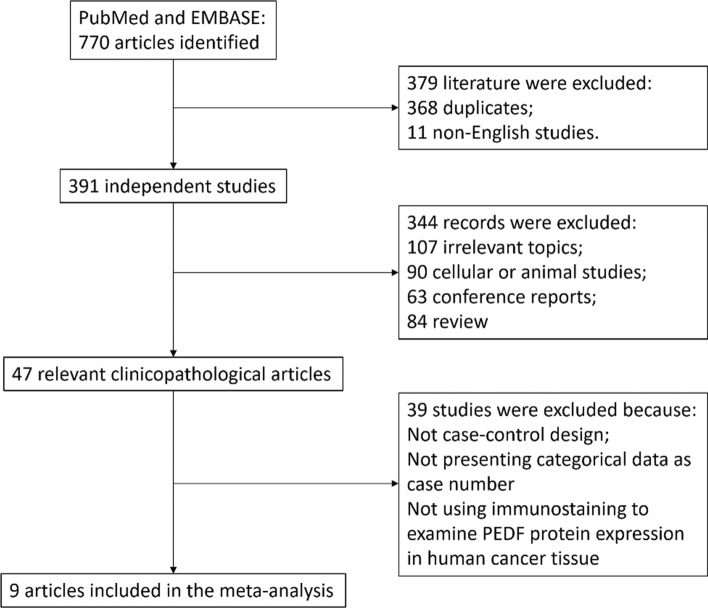
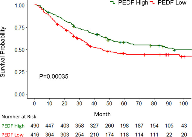
Methods: We performed a comprehensive search for the studies on PEDF expression in 14 top-ranked types of solid tumor cancer with the highest incidence. A systemic approach was used to screen for qualified studies and to extract data. Meta-analysis was performed to investigate if PEDF expression is associated with the TNM staging, tumor size, lymph node invasion, distal metastasis and pathological grade of tumor in a pan-cancer manner. A Kaplan-Meier curve was plotted with the digitally-reconstituted patient survival data to study the effect of PEDF expression on the prognosis of cancer patients.

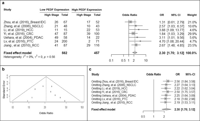
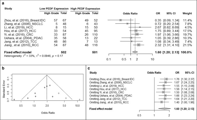
Results: A total of nine studies were selected, reviewed and analyzed. Meta-analysis suggested that decreased PEDF protein expression was associated with higher TNM staging (OR = 2.13, 95% CI: 1.61-2.81), larger tumor size (OR = 1.42, 95% CI: 1.1-1.84), larger possibility of lymph node invasion (OR = 1.68, 95% CI: 1.26-2.22) and higher pathological grade (OR = 1.6, 95% CI: 1.2-2.13). No correlation was found between PEDF expression and tumor distal metastasis, gender or age. In addition, low PEDF protein level in tumor tissue is correlated with shorter overall survival (P < 0.05).

Conclusions: Low PEDF protein expression in cancer is significantly associated with more advanced cancer progression and significantly poorer survival. The differential clinical outcome among patients with various PEDF expression suggests its prognostic value.

Keywords: Cancer progression; Meta-analysis; Overall survival; Pigment epithelium derived factor (PEDF); Prognosis.


Conflict of interest statement

The authors declare that they have no conflicts of interest to declare that are relevant to the content of this article.

Figures

Fig. 1
Fig. 1
Flow diagram and results of literature review. The flow diagram depicts the step-wise screening of the retrieved articles from database, including the number and reasons of exclusion. A total of 9 studies were selected by this unbiased process
Fig. 2
Fig. 2
Cancer with low PEDF expression has higher odds to have higher TNM staging. a Forest plot showing the association of PEDF expression and cancer TNM staging. Squares indicate study-specific odds ratios (ORs). The size of the box is proportional to the weight of the study. Horizontal lines indicate 95% confidence intervals (CI). A diamond indicates the summary OR with its corresponding 95% CI. b Funnel plot on the publication bias of studies on PEDF expression and TNM staging. Each filled circle represents one study. Open circles represent added artificial studies to correct publication bias. c Forest plot of sensitivity test. Squares indicate the summary OR after omitting one specific study. Horizontal lines indicate 95% CI. A diamond indicates the summary OR of all the studies with its corresponding 95% CI. IDC intra-ductal carcinoma, NSCLC non-small cell lung cancer, HCC hepatocellular carcinoma, CRC colorectal carcinoma, PDAC pancreatic ductal adenocarcinoma, PTC papillary thyroid carcinoma, RCC renal cell carcinoma
Fig. 3
Fig. 3
Cancer with low PEDF protein expression is larger in size. a Forest plot showing the association of PEDF expression and tumor size. Squares indicate study-specific odds ratios (ORs). The size of the box is proportional to the weight of the study. Horizontal lines indicate 95% confidence interval (CI). A diamond indicates the summary OR with its corresponding 95% CI. b Funnel plot showing no publication bias of studies on PEDF expression and tumor size. Each filled circle represents one study. c Forest plot of sensitivity test. Squares indicate the summary OR after omitting one specific study. Horizontal lines indicate 95% CI. A diamond indicates the summary OR of all the studies with its corresponding 95% CI. IDC intra-ductal carcinoma, NSCLC non-small cell lung cancer, HCC hepatocellular carcinoma, CRC colorectal carcinoma, PDAC pancreatic ductal adenocarcinoma, PTC papillary thyroid carcinoma, RCC renal cell carcinoma
Fig. 4
Fig. 4
Cancer with low PEDF protein expression has higher odds to invade lymph nodes. a Forest plot showing the association of PEDF expression and lymph node invasion. Squares indicate study-specific odds ratios (ORs). The size of the box is proportional to the weight of the study. Horizontal lines indicate 95% confidence interval (CI). A diamond indicates the summary OR with its corresponding 95% CI. b Funnel plot on the publication bias of studies on PEDF expression and lymph node invasion. Each filled circle represents one study. Open circles represent filled studies to correct publication bias. c Forest plot of sensitivity test. Squares indicate the summary OR after omitting one specific study. Horizontal lines indicate 95% CI. A diamond indicates the summary OR of all the studies with its corresponding 95% CI. +LN positive lymph node, IDC intra-ductal carcinoma, NSCLC non-small cell lung cancer, HCC hepatocellular carcinoma, CRC colorectal carcinoma, PDAC pancreatic ductal adenocarcinoma, PTC papillary thyroid carcinoma, RCC renal cell carcinoma
Fig. 5
Fig. 5
Cancer with low PEDF expression has increased odds to have higher pathological grade. a Forest plot showing the association of PEDF expression and tumor pathological grade. Squares indicate study-specific odds ratios (ORs). The size of the box is proportional to the weight of the study. Horizontal lines indicate 95% confidence interval (CI). A diamond indicates the summary OR with its corresponding 95% CI. b Funnel plot on the publication bias of studies on PEDF expression and tumor pathological grade. Each filled circle represents one study. Open circles represent filled studies to correct publication bias. c Forest plot of sensitivity test. Squares indicate the summary OR after omitting one specific study. Horizontal lines indicate 95% CI. A diamond indicates the summary OR of all the studies with its corresponding 95% CI. IDC intra-ductal carcinoma, NSCLC non-small cell lung cancer, HCC hepatocellular carcinoma, CRC colorectal carcinoma, PDAC pancreatic ductal adenocarcinoma, TCC (bladder) transitional cell carcinoma, RCC renal cell carcinoma
Fig. 6
Fig. 6
Cancer patients with high-PEDF protein expression have longer overall survival shown in Kaplan–Meier curves. Green line represents the OS of patients with high PEDF-expressing cancers whereas red line represents the OS of patients with low PEDF-expressing cancers. Below the graph shows the number of patients at risk for each group at a particular time point

Similar articles

Cited by

References

    1. Tombran-Tink J, Johnson LV. Neuronal differentiation of retinoblastoma cells induced by medium conditioned by human RPE cells. Invest Ophthalmol Vis Sci. 1989;30(8):1700–1707. - PubMed
    1. Becerra SP, Palmer I, Kumar A, Steele F, Shiloach J, Notario V, Chader GJ. Overexpression of fetal human pigment epithelium-derived factor in Escherichia coli. A functionally active neurotrophic factor. J Biol Chem. 1993;268(31):23148–23156. - PubMed
    1. Carmeliet P, Jain RK. Angiogenesis in cancer and other diseases. Nature. 2000;407(6801):249–257. - PubMed
    1. Folkman J, Bach M, Rowe JW, Davidoff F, Lambert P, Hirsch C, Goldberg A, Hiatt HH, Glass J, Henshaw E. Tumor angiogenesis—therapeutic implications. New Engl J Med. 1971;285(21):1182–2000. - PubMed
    1. Dawson DW, Volpert OV, Gillis P, Crawford SE, Xu H, Benedict W, Bouck NP. Pigment epithelium-derived factor: a potent inhibitor of angiogenesis. Science. 1999;285(5425):245–248. - PubMed

LinkOut - more resources