. 2022 Feb 3;13(1):9.
doi: 10.1007/s12672-022-00470-9.

Dual inhibition of anti-apoptotic proteins BCL-XL and MCL-1 enhances cytotoxicity of Nasopharyngeal carcinoma cells

Affiliations

Dual inhibition of anti-apoptotic proteins BCL-XL and MCL-1 enhances cytotoxicity of Nasopharyngeal carcinoma cells

Siti Fairus Abdul Rahman et al. Discov Oncol. .

Abstract

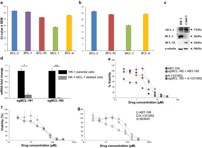
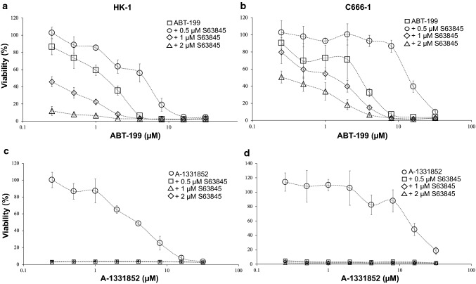
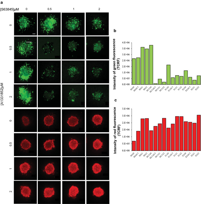
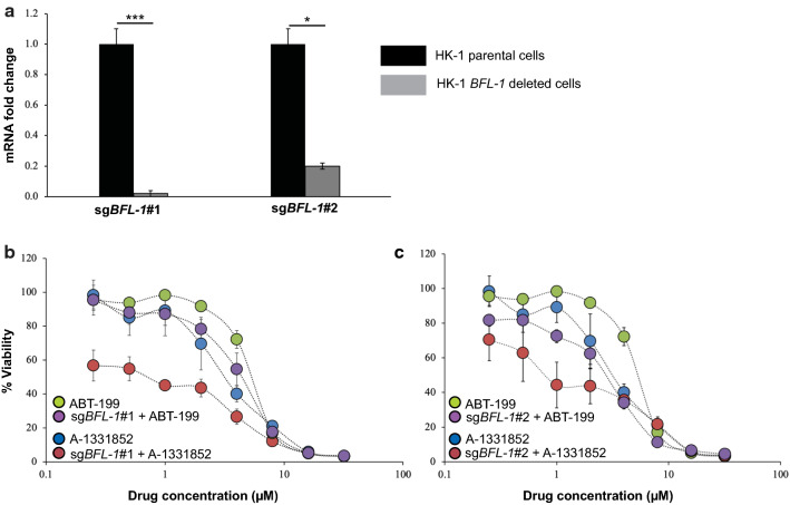
One of the many strategies that cancer cells evade death is through up-regulation of the BCL-2 anti-apoptotic proteins. Hence, these proteins have become attractive therapeutic targets. Given that different cell populations rely on different anti-apoptotic proteins for survival, it is crucial to determine which proteins are important for Nasopharyngeal carcinoma (NPC) cell survival. Here we determined the survival requirements for the NPC cells using a combination of the CRISPR/Cas9 technique and selective BH3-mimetics. A human apoptosis RT2 Profiler PCR Array was first employed to profile the anti-apoptotic gene expressions in NPC cell lines HK-1 and C666-1. The HK-1 cells expressed all the anti-apoptotic genes (MCL-1, BFL-1, BCL-2, BCL-XL, and BCL-w). Similarly, the C666-1 cells expressed all the anti-apoptotic genes except BFL-1 (undetectable level). Notably, both cell lines highly expressed MCL-1. Deletion of MCL-1 sensitized the NPC cells to BCL-XL selective inhibitor A-1331852, suggesting that MCL-1 and BCL-XL may be important for NPC cell survival. Co-inhibition of MCL-1 and BCL-2 with MCL-1 selective inhibitor S63845 and BCL-2 selective inhibitor ABT-199 inhibited NPC cell proliferation but the effect on cell viability was more profound with co-inhibition of MCL-1 and BCL-XL with S63845 and A-1331852, implying that MCL-1 and BCL-XL are crucial for NPC cell survival. Furthermore, co-inhibition of MCL-1 and BCL-XL inhibited the growth and invasion of NPC spheroids. Deletion of BFL-1 sensitized NPC cells to A-1331852 suggesting that BFL-1 may play a role in NPC cell survival. Taken together co-inhibition of BCL-XL and MCL-1/BFL-1 could be potential treatment strategies for NPC.

Keywords: BCL-XL; BFL-1; BH3 mimetics; MCL-1; Nasopharyngeal carcinoma; Spheroids.


Conflict of interest statement

The authors declare no conflict of interest.

Figures

Fig. 1
Fig. 1
HK-1 MCL-1 deleted cells were sensitive to treatment of BCL-XL selective inhibitor A-1331852. Expression of the anti-apoptotic genes was determined using the human apoptosis real-time PCR array (RT2 Profiler PCR Array). Ct values are inversely proportional to the amount of target gene present in the cells. The a HK-1 expressed all of the anti-apoptotic genes and b C666-1 expressed all of the anti-apoptotic genes, except BFL-1. Both cell lines expressed high levels of MCL-1. Error bars show the standard error of the mean (SEM). c Basal expression levels of the anti-apoptotic protein in the NPC cell lines. The basal expression levels of MCL-1, BCL-2, and BCL-XL in the C666-1 and the HK-1 cells were determined by SDS-PAGE gel electrophoresis (this image was taken from [10] with permission). d qPCR validation MCL-1 gene deletion in the HK-1 cells. MCL-1 expression levels were normalized to parental cells. Bars indicate the mean SEM of three independent experiments. Statistically significant differences in MCL-1 expression between the parental cell line and the MCL-1 knockout cells are shown as ***p ≤ 0.001 or *p < 0.05 determined by a two-tailed paired T-test. e The sensitivity of the HK-1 parental cell line and the HK-1 sgMCL-1#2 cells were tested to the single-agent treatment of either ABT-199 or A-1331852 (0–32 μM). Points represent the SD of four experiments. f HK-1 and g C666-1 were treated with increasing concentrations of either ABT-199, A-1331852, or S63845 (0–32 μM). Points represent the SD of four experiments
Fig. 2
Fig. 2
Co-inhibition of BCL-2 and MCL-1 using ABT-199 and S63845. a HK-1 cells were treated with increasing concentrations of ABT-199 (0–32 μM) in the presence and absence of S63845. Points represent the SD of four experiments. b C666-1 cells were treated with increasing concentrations of ABT-199 (0–32 μM) in the presence and absence of S63845. Points represent the SD of four experiments. Co-inhibition of BCL-XL and MCL-1 using A-1331852 and S63845. c HK-1 and d C666-1 cells were treated with increasing concentrations of A-1331852 (0–32 μM) in the presence and absence of S63845. Points represent the SD of four experiments
Fig. 3
Fig. 3
The effect of the combination of S63845 and A-1331852 on the growth and invasion of HK-1 spheroids over 3 days. a The spheroids were treated with single agents S63845 and A-1331852 and a combination of both over three days at the indicated concentrations, n = 2–3 spheroids per combination. Cell viability was determined using the live/dead assay (Viable cells: stained green by Calcein-AM; Dead cells: stained red by Ethidium-homodimer I). Size bar: 200 μm. The intensity of b green and c red fluorescence was measured for each drug combination and presented as TCRF, n = 2 spheroids per combination. TCRF: Total Corrected Red Fluorescence; “S” denotes S63845 and “A” denotes A-1331852. The numbers next to “S” and “A” indicate the doses of the drug combinations
Fig. 4
Fig. 4
HK-1 BFL-1 deleted cells were sensitive to treatment of BCL-XL selective inhibitor A-1331852. a Quantitative PCR (qPCR) validation BFL-1 gene deletion in the HK-1 cells. BFL-1 expression levels were normalized to parental cells. Bars indicate the SD of three independent experiments. Statistically significant differences in BFL-1 expression between the parental cell line and the BFL-1 knockout cells are shown as ***p ≤ 0.001 or *p < 0.05 determined by a two-tailed paired T-test. The sensitivity of the HK-1 parental cell line and the b HK-1 sgBFL-1#1 and c HK-1 sgBFL-1#2 cells were tested to single-agent treatment of either ABT-199 or A-1331852 (0–32 μM). Points represent the SD of four experiments

Similar articles

Cited by

References

    1. Global Cancer Observatory: Cancer Today. Lyon, France: International Agency for Research on Cancer (IARC). https://gco.iarc.fr/today. Accessed 2 Nov 2021.
    1. Al-Sarraf M, LeBlanc M, Giri PG, et al. Chemoradiotherapy versus radiotherapy in patients with advanced nasopharyngeal cancer: phase III randomized Intergroup study 0099. J Clin Oncol. 1998 doi: 10.1200/jco.1998.16.4.1310. - DOI - PubMed
    1. Kurschat C, Metz A, Kirschnek S, et al. Importance of Bcl-2-family proteins in murine hematopoietic progenitor and early B cells. Cell Death Dis. 2021 doi: 10.1038/s41419-021-04079-8. - DOI - PMC - PubMed
    1. Sheu LF, Chen A, Meng CL, et al. Analysis of bcl-2 expression in normal, inflamed, dysplastic nasopharyngeal epithelia, and nasopharyngeal carcinoma: association with p53 expression. Hum Pathol. 1997 doi: 10.1016/s0046-8177(97)90078-2. - DOI - PubMed
    1. Yu Y, Dong W, Li X, et al. Significance of c-Myc and Bcl-2 protein expression in nasopharyngeal carcinoma. Arch Otolaryngol Head Neck Surg. 2003 doi: 10.1001/archotol.129.12.1322. - DOI - PubMed

LinkOut - more resources