

Blood Compatibility of Hydrophilic Polyphosphoesters
- PMID: 35201742
- PMCID: PMC8941511
- DOI: 10.1021/acsabm.1c01210
Blood Compatibility of Hydrophilic Polyphosphoesters
Abstract
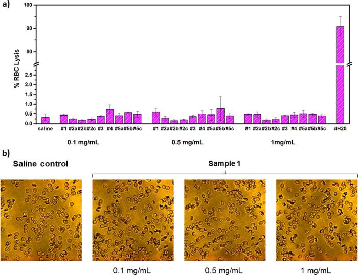
Polyphosphoesters (PPEs) are a class of versatile degradable polymers. Despite the high potential of this class of polymers in biomedical applications, little is known about their blood interaction and compatibility. We evaluated the hemocompatibility of water-soluble PPEs (with different hydrophilicities and molar masses) and PPE-coated model nanocarriers. Overall, we identified high hemocompatibility of PPEs, comparable to poly(ethylene glycol) (PEG), currently used for many applications in nanomedicine. Hydrophilic PPEs caused no significant changes in blood coagulation, negligible platelet activation, the absence of red blood cells lysis, or aggregation. However, when a more hydrophobic copolymer was studied, some changes in the whole blood clot strength at the highest concentration were detected, but only concentrations above that are typically used for biomedical applications. Also, the PPE-coated model nanocarriers showed high hemocompatibility. These results contribute to defining hydrophilic PPEs as a promising platform for degradable and biocompatible materials in the biomedical field.
Keywords: RBC interaction; biodegradable polymers; blood coagulation; hemocompatibility; platelet activation; poly(ethylene glycol); polyphosphoesters.
Conflict of interest statement
The authors declare no competing financial interest.
Figures


References
-
- Pelosi C.; Tinè M. R.; Wurm F. R. Main-Chain Water-Soluble Polyphosphoesters: Multi-Functional Polymers as Degradable PEG-Alternatives for Biomedical Applications. Eur. Polym. J. 2020, 141, 110079 10.1016/j.eurpolymj.2020.110079. - DOI
-
- Bauer K. N.; Tee H. T.; Velencoso M. M.; Wurm F. R. Main-Chain Poly(Phosphoester)s: History, Syntheses, Degradation, Bio-and Flame-Retardant Applications. Prog. Polym. Sci. 2017, 73, 61–122. 10.1016/j.progpolymsci.2017.05.004. - DOI
-
- Bauer K. N.; Liu L.; Wagner M.; Andrienko D.; Wurm F. R. Mechanistic Study on the Hydrolytic Degradation of Polyphosphates. Eur. Polym. J. 2018, 108, 286–294. 10.1016/j.eurpolymj.2018.08.058. - DOI
-
- Wang F.; Wang Y. C.; Yan L. F.; Wang J. Biodegradable Vesicular Nanocarriers Based on Poly(ε-Caprolactone)-Block-Poly(Ethyl Ethylene Phosphate) for Drug Delivery. Polymer 2009, 50, 5048–5054. 10.1016/j.polymer.2009.09.007. - DOI
Publication types
MeSH terms
Substances
Grants and funding
LinkOut - more resources
Full Text Sources
