. 2022 Feb 25;375(6583):eabh3021.
doi: 10.1126/science.abh3021. Epub 2022 Feb 25.

Hyperexcitable arousal circuits drive sleep instability during aging

Affiliations

Hyperexcitable arousal circuits drive sleep instability during aging

Shi-Bin Li et al. Science. .

Abstract

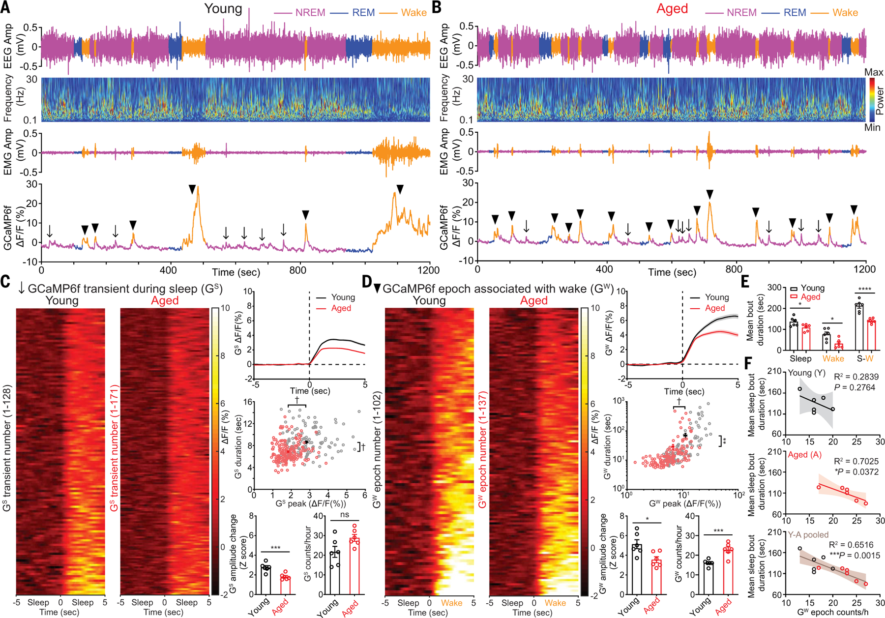
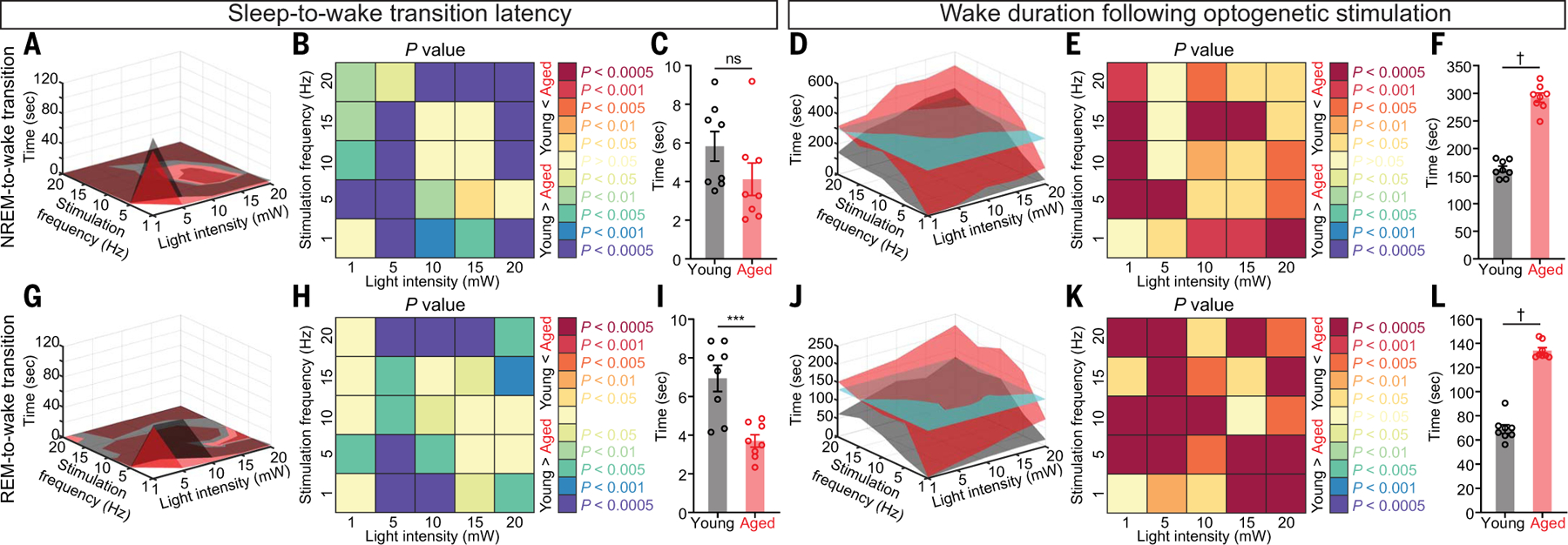
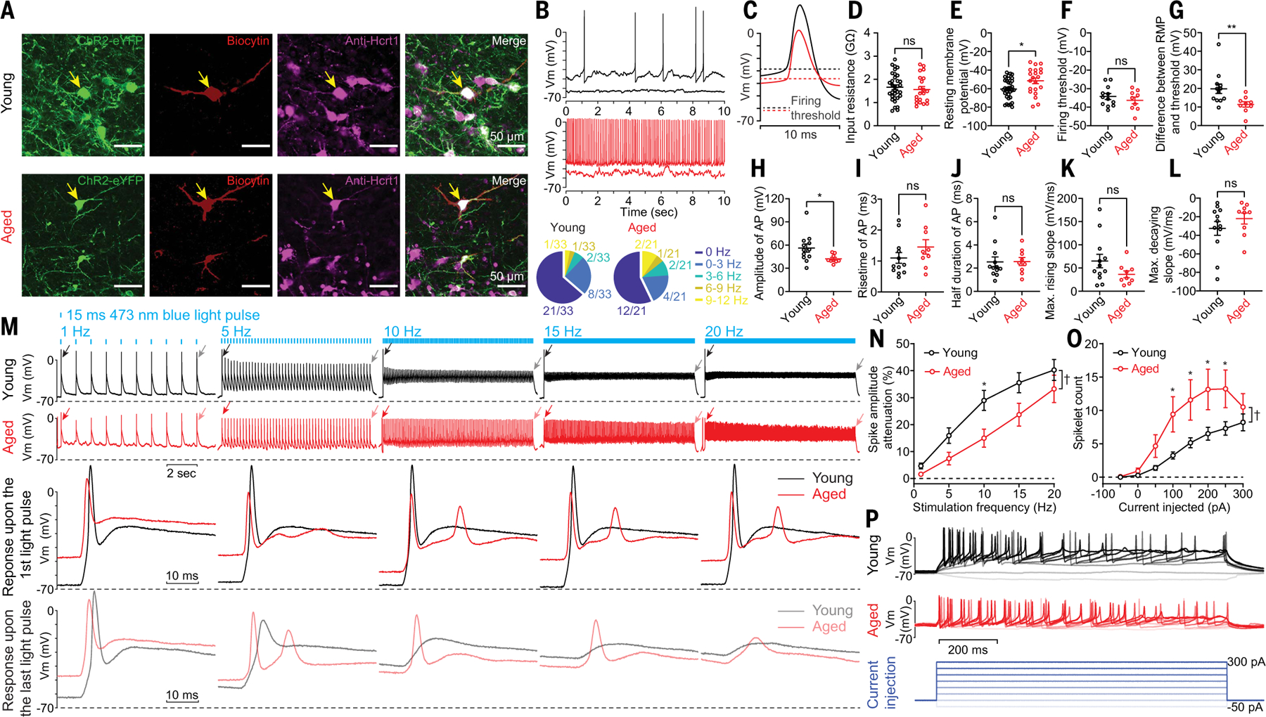
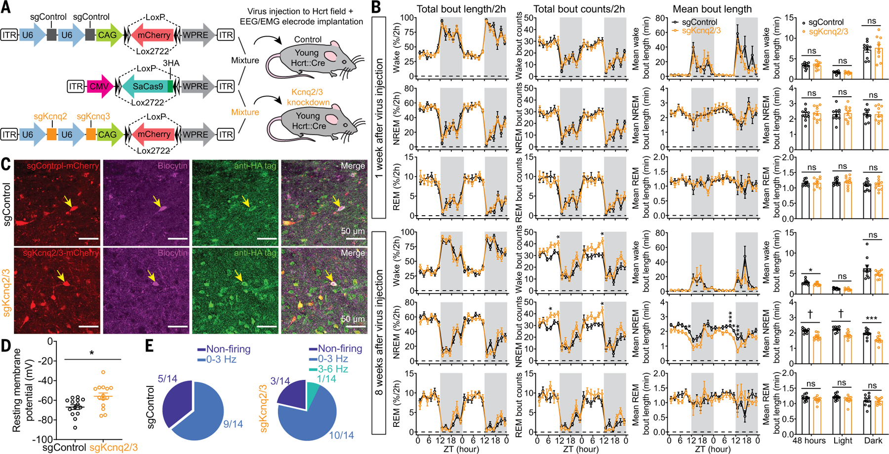
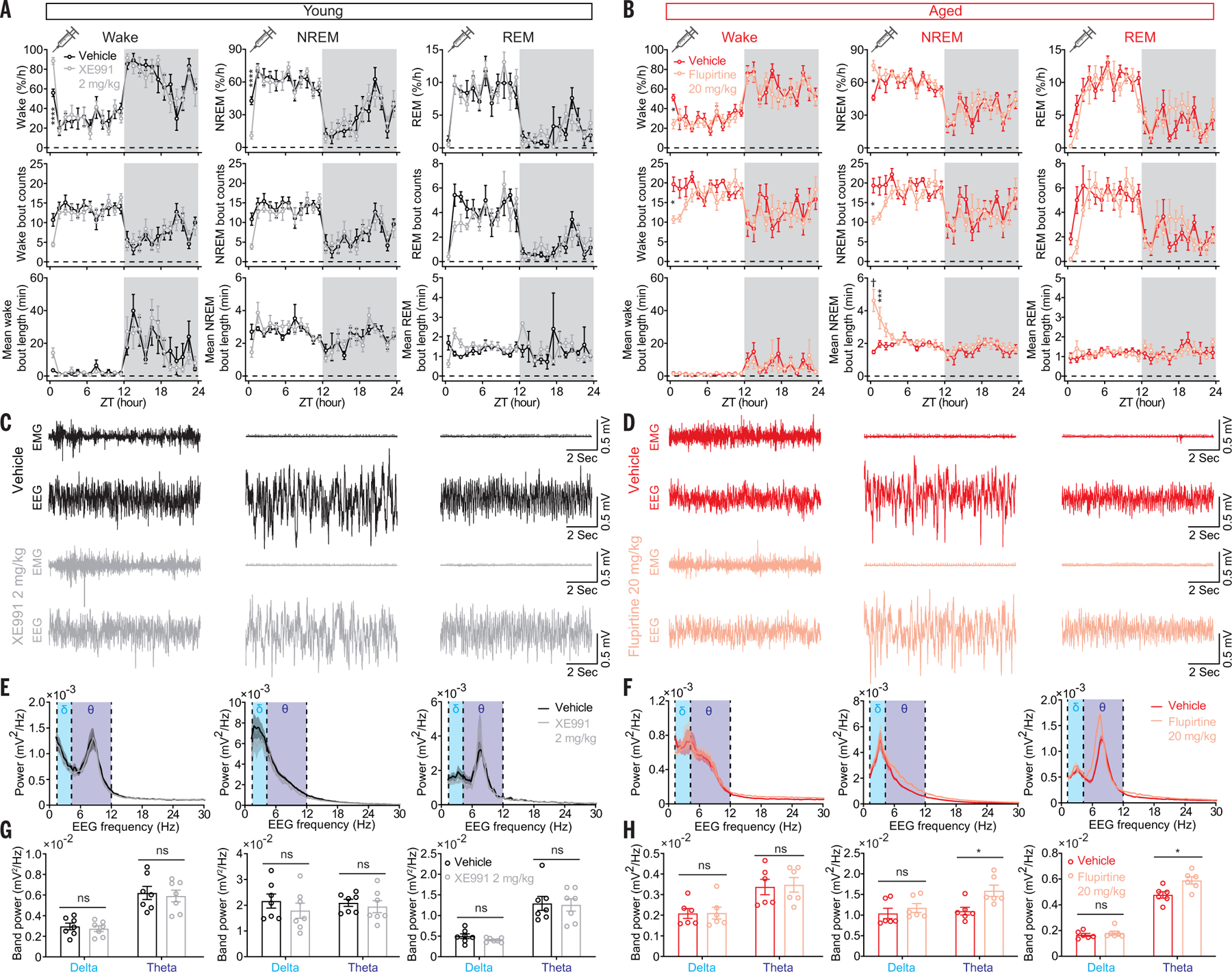
Sleep quality declines with age; however, the underlying mechanisms remain elusive. We found that hyperexcitable hypocretin/orexin (Hcrt/OX) neurons drive sleep fragmentation during aging. In aged mice, Hcrt neurons exhibited more frequent neuronal activity epochs driving wake bouts, and optogenetic activation of Hcrt neurons elicited more prolonged wakefulness. Aged Hcrt neurons showed hyperexcitability with lower KCNQ2 expression and impaired M-current, mediated by KCNQ2/3 channels. Single-nucleus RNA-sequencing revealed adaptive changes to Hcrt neuron loss in the aging brain. Disruption of Kcnq2/3 genes in Hcrt neurons of young mice destabilized sleep, mimicking aging-associated sleep fragmentation, whereas the KCNQ-selective activator flupirtine hyperpolarized Hcrt neurons and rejuvenated sleep architecture in aged mice. Our findings demonstrate a mechanism underlying sleep instability during aging and a strategy to improve sleep continuity.


Conflict of interest statement

Competing interests: L.d.L. and S.-B.L. are co-inventors on a WIPO (World Intellectual Property Organization) patent WO/2021/119018 related to the work described in this manuscript. L.d.L. is a Chief Scientific Advisor to and receives consulting fees from Attune Neurosciences. L.d.L. is a paid consultant to Eisai Pharmaceuticals and Idorsia Pharmaceuticals. G.S. is a cofounder of Epiodyne, a drug discovery company, an inventor on a patent application (US application no. 62/962,581) related to imaging of neural dynamics to discover analgesics, and a member of the NIH PSPP (Preclinical Screening Platform for Pain) External Consulting Board. The remaining authors declare that they have no competing interests.

Figures

Fig. 1.
Fig. 1.. Spontaneous activity of Hcrt neurons across sleep/wake states in young and aged mice.
(A and B) Representative EEG, EEG power spectra, EMG, simultaneous Hcrt GCaMP6f signals from (A) young and (B) aged mice. The arrows indicate GCaMP6f transients during sleep (GS), and the black triangles indicate GCaMP6f epochs associated with wakefulness (GW). (C) Staged GS signals during 10 s around the start of GS transients from identical length (6 hours, 1 hour/mouse) of recorded GCaMP6f signals from young and aged mice (n = 6 mice each group), respectively, during light phase, averaged trace plot (right top), scatter plot of individual GS duration against GS peak (young, n = 128; aged, n = 171) (right middle), animal-based comparison of GS signals for Z score and GS frequency (right bottom). (D) Staged GW signals during 10 s around start of GW epochs from identical length (6 hours, 1 hour/mouse) of recorded GCaMP6f signals from young and aged mice, averaged trace plot (right top), scatter plot of individual GW duration against GW peak (young, n = 102; aged, n = 137) (right middle), animal-based comparison of GW signals for Z score and GW frequency (right bottom). (E) Animal-based comparison of mean bout duration for sleep, wake, and entire S-W episodes (n = 6 mice each group). (F) Correlation for mean sleep bout duration against GW bout counts/hour in young, aged, and pooled datasets. Data represent mean ± SEM. In (C) to (E) unpaired t test with Welch’s correction; (F), Spearman correlation, linear fit and 95% confidence band; *P < 0.05, **P < 0.01, ***P < 0.005, ****P < 0.001, †P < 0.0005; statistical details see supplementary text.
Fig. 2.
Fig. 2.. More prolonged wake bouts upon optogenetic stimulation of Hcrt neurons expressing ChR2-eYFP in aged mice.
(A) Surface plot of NREM-to-wake transition latency based on the mean value of each stimulation condition. (B and C) Comparison of NREM-to-wake transition latency based on (B) each stimulation condition and (C) the mean value for each animal. (D) Surface plot of wake duration based on the mean value of each stimulation condition. The cyan cutaway surface indicates the mean value for the aged group. (E and F) Comparison of wake duration based on (E) each stimulation condition and (F) the mean value for each animal. (G) Surface plot of REM-to-wake transition latency based on the mean value of each stimulation condition. (H and I) Comparison of REM-to-wake transition latency based on (H) each stimulation condition and (I) the mean value for each animal. (J) Surface plot of wake duration based on the mean value of each stimulation condition. The cyan cutaway surface indicates the mean value for the aged group. (K and L) Comparison of wake duration based on (K) each stimulation condition and (L) the mean value for each animal. In (B), (C), (E), (F), (H), (I), (K), and (L): Mann-Whitney U test; ***P < 0.005, †P < 0.0005. Statistical details are available in the supplementary text.
Fig. 3.
Fig. 3.. Hyperexcitability in aged Hcrt neurons revealed with whole-cell patch clamp recording.
(A) Representative slices containing recorded ChR2-eYFP–labeled Hcrt neurons infused with biocytin. (B) Representative traces and fractions of young and aged Hcrt neurons with and without spontaneous firing activities. (C) Averaged traces of spontaneous APs shown in (B). (D to L) Comparison of basic electrophysiological properties between young and aged Hcrt neurons. (D) Input resistance and (E) RMP of all the recorded young and aged Hcrt neurons (young, n = 33 neurons versus aged, n = 21 neurons from eight mice each group). Comparison of other parameters including (F) firing threshold, (G) difference between RMP and firing threshold, (H) AP peak amplitude, (I) AP rising time, (J) AP half duration, (K) maximum rising slope, and (L) decaying slope of spontaneous APs between young and aged Hcrt neurons (young, n = 12 neurons versus aged, n = 9 neurons from eight mice each group). (M) (Top) Representative traces of young and aged Hcrt neurons expressing ChR2-eYFP upon optogenetic stimulation. (Bottom) The same responses on a slower time base, illustrating the response to the first and last light pulse stimulations at each stimulation frequency (membrane voltage/Vm). (N) Significant reduction of response attenuation calculated based on the first and the last response from trains as in M (young, n = 23 neurons versus aged, n = 21 neurons from eight mice each group). (O and P) Step current injections triggered more spikelets in aged Hcrt neurons than in young Hcrt neurons [(O) young, n = 33 neurons versus aged, n = 26 neurons from eight mice each group; (P) representative traces and current injection protocol]. In (D) to (L): Mann-Whitney U test; (N) and (O): two-way ANOVA followed by post hoc Šidák’s multiple comparisons; *P < 0.05, **P < 0.01, †P < 0.0005. Statistical details are available in the supplementary text.
Fig. 4.
Fig. 4.. Impaired IM associated with loss of KCNQ2 in aged Hcrt neurons.
(A) Representative traces from a young Hcrt neuron (left) before or (right) in the presence of a KCNQ2/3 blocker XE991 (50 μM). (B) XE991 depolarized RMP and (C) increased firing frequency in young Hcrt neurons (n = 19 neurons from seven mice). (D) Representative traces from an aged Hcrt neuron (left) before or (right) in the presence of a KCNQ2/3 activator flupirtine (50 μM). (E) Flupirtine hyperpolarized RMP and (F) decreased firing frequency in aged Hcrt neurons (n = 8 neurons from five mice). (G) IM in young Hcrt neurons modulated by XE991 (top; n = 6 neurons from three mice) and flupirtine (bottom; n = 10 neurons from five mice). (H) IM in aged Hcrt neurons modulated by XE991 (top; n = 7 neurons from three mice) and flupirtine (bottom; n = 15 neurons from five mice). (I) Basal IM in young Hcrt neurons (n = 25 neurons from nine mice) versus in aged Hcrt neurons (n = 26 neurons from nine mice). (J) Array tomography revealed reduced KCNQ2 expression in aged Hcrt neurons (n = 4 mice/group). In (B) and (C), Wilcoxon matched-pairs signed rank test; (E) and (F), RM one-way ANOVA followed by post hoc Tukey’s multiple comparisons; (G), (H), and (J), paired t test; (I) unpaired t test with Welch’s correction; statistical details are available in the supplementary text.
Fig. 5.
Fig. 5.. CRISPR/SaCas9–mediated disruption of Kcnq2/3 genes in Hcrt neurons leads to NREM sleep fragmentation in young mice.
(A) Schematic of AAV sgControl, AAV SaCas9, AAV sgKcnq2/3 vector design, and bilateral viral infection of Hcrt neurons in young Hcrt∷Cre mice. (B) Two-hour (left) binned percentage, (middle left) bout counts, (middle right) mean bout length and (right) mean bout length based on circadian phase for wake, NREM, and REM sleep at 1 week (top) and 8 weeks (bottom) after injection of a virus mixture, as illustrated in (A) (n = 10 mice/group, dark phase indicated by gray shielding). (C) Representative slices expressing sgRNA with fluorescent mCherry flag and SaCas9–3HA for sgControl and sgKcnq2/3 group, respectively. Patch clamp recorded cells were labeled with biocytin, and post hoc antibody staining against HA tag confirmed the cells expressing SaCas9 for data analyses. (D) Comparison of RMPs between sgControl and sgKcnq2/3 group (n = 14 neurons from three mice each group). (E) Fractions of neurons with different firing frequencies in the (left) sgControl and (right) sgKcnq2/3 groups. Data indicate mean ± SEM [(B) left to middle right, two-way RM ANOVA followed by Šidák’s multiple comparisons; (B) right, Holm-Šidák; (D) Mann-Whitney U test; *P < 0.05, ***P < 0.005, †P < 0.0005; statistical details are avialable in the supplementary text].
Fig. 6.
Fig. 6.. Pharmacological manipulation of sleep/wake states with KCNQ2/3 ligands.
(A) Significantly increased wake amount by the KCNQ2/3 blocker XE991 (2 mg/kg) in young mice. (B) Significantly increased NREM amount and mean bout length by the KCNQ2/3 activator flupirtine (20 mg/kg) in aged mice. (C) Representative EMG-EEG raw traces from vehicle- and XE991-treated (2 mg/kg) young mice. (D) Representative EMG-EEG raw traces from vehicle- and flupirtine-treated (20 mg/kg) aged mice. (E) Power spectra of EEG for vehicle- and XE991-treated young mice and (F) power spectra of EEG for vehicle- and flupirtine-treated aged mice. (G) Comparison of delta, theta band power between vehicle- and XE991-treated young mice and (H) between vehicle- and flupirtine-treated aged mice. Data indicate mean ± SEM [young, n = 7 mice each group; aged, n = 6 mice each group; (A) and (B) two-way liner mixed-effects model followed by Šidák’s multiple comparisons, dark phase indicated by gray shielding; (G) and (H) Holm-Šidák, *P < 0.05, ***P < 0.005, ****P < 0.001, †P < 0.0005; statistical details are available in the supplementary text].

Comment in

References

    1. Scullin MK, Bliwise DL, Sleep, cognition, and normal aging: Integrating a half century of multidisciplinary research. Perspect. Psychol. Sci 10, 97–137 (2015). doi: 10.1177/1745691614556680; - DOI - PMC - PubMed
    1. Yaffe K, Falvey CM, Hoang T, Connections between sleep and cognition in older adults. Lancet Neurol 13, 1017–1028 (2014). doi: 10.1016/S1474-4422(14)70172-3; - DOI - PubMed
    1. Carskadon MA, Brown ED, Dement WC, Sleep fragmentation in the elderly: Relationship to daytime sleep tendency. Neurobiol. Aging 3, 321–327 (1982). doi: 10.1016/0197-4580(82)90020-3; - DOI - PubMed
    1. Mander BA, Winer JR, Walker MP, Sleep and human aging. Neuron 94, 19–36 (2017). doi: 10.1016/j.neuron.2017.02.004; - DOI - PMC - PubMed
    1. Rodriguez JC, Dzierzewski JM, Alessi CA, Sleep problems in the elderly. Med. Clin. North Am 99, 431–439 (2015). doi: 10.1016/j.mcna.2014.11.013; - DOI - PMC - PubMed

Publication types

MeSH terms