. 2022 Mar 11;375(6585):1173-1176.
doi: 10.1126/science.abn6840. Epub 2022 Feb 24.

Structural basis of human telomerase recruitment by TPP1-POT1

Affiliations

Structural basis of human telomerase recruitment by TPP1-POT1

Zala Sekne et al. Science. .

Abstract

Telomerase maintains genome stability by extending the 3' telomeric repeats at eukaryotic chromosome ends, thereby counterbalancing progressive loss caused by incomplete genome replication. In mammals, telomerase recruitment to telomeres is mediated by TPP1, which assembles as a heterodimer with POT1. We report structures of DNA-bound telomerase in complex with TPP1 and with TPP1-POT1 at 3.2- and 3.9-angstrom resolution, respectively. Our structures define interactions between telomerase and TPP1-POT1 that are crucial for telomerase recruitment to telomeres. The presence of TPP1-POT1 stabilizes the DNA, revealing an unexpected path by which DNA exits the telomerase active site and a DNA anchor site on telomerase that is important for telomerase processivity. Our findings rationalize extensive prior genetic and biochemical findings and provide a framework for future mechanistic work on telomerase regulation.


Conflict of interest statement

Competing interests: Authors declare that they have no competing interests.

Figures

Fig. 1
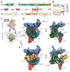
Fig. 1. Structures of telomerase catalytic core with telomeric DNA and shelterin components.
(A) Domain organization of protein subunits in the catalytic core and TPT complex. Regions not observed in the structures are displayed as semi-transparent. Domain colors are used thorough unless indicated otherwise. (B) Secondary structure of hTR. (C) Schematic of human telomerase bound to TPP1 and POT1. (D) 3.2 Å cryo-EM reconstruction of telomerase catalytic core-DNA-TPP1 complex (fig. S6). (E) 3.9 Å cryo-EM reconstruction of telomerase catalytic core-DNA-TPP1-POT1 complex (fig. S7).
Fig. 2
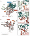
Fig. 2. Telomerase-TPP1 interactions.
(A) Interactions between TPP1 OB-fold domain and TERT. Circles indicate the three contacts that the NOB (orange) and TEL-patch (red) of TPP1 make with TERT. The β-hairpin, which become ordered upon TPP1 binding, is highlighted. (B) Comparison of the unbound (grey, PDB 2I46 (16)) and telomerase-bound (colored) OB-fold domain of TPP1. (C) Close-up view of the interactions between the NOB of TPP1 and TERT (fig. S6I). (D and E) Close-up views of the interactions between the TEL-patch and TERT (fig. S6, J and K).
Fig. 3
Fig. 3. Telomerase-POT1 interactions.
(A) Structure of telomerase-DNA-TPP1-POT1 complex. (B) 3.9 Å cryo-EM reconstruction of telomerase-DNA-TPP1-POT1 complex. The inset highlights interactions between POT1 and TERT. The N-DAT region (purple) interacts with both TPP1 and POT1.
Fig. 4
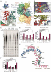
Fig. 4. Telomeric DNA substrate.
(A) The DNA path guided by TERT, hTR and POT1. The left upper panel shows how the view is related to the telomerase-DNA-TPP1-POT1 reconstruction. (B) Interactions between the template RNA and the DNA upstream of the RNA template-DNA duplex (fig. S6L). (C) Close-up view of the interactions between the DNA substrate and the proposed anchor site on the TEN domain and the IFD (fig. S6N). (D) Interactions between the flipped-out G23 DNA base and TERT (fig. S6M). (E) Telomerase activity assays in the absence and presence of the TPT complex for wild-type (WT) enzyme and TERT mutants at the proposed DNA anchor site (Y176A/Q177A, K757A/F759A and Q794A) (fig. S6, H and N). Assays were performed after enzyme enrichment on MagStrepXT resins using ZZ-TEV-twin-Strep tagged TERT. RC, recovery control. Experiments were performed in triplicate. Detailed analyses of the input lysates are shown in fig. S12. (F and G) Bar graphs showing the quantifications of telomerase activity and processivity of the activity assays shown in (E), respectively. Values were normalized to telomerase without TPT. Error bars are the standard errors of the mean (SEM) obtained from the replicates. (H) Schematic of the DNA substrate and its interactions with TERT, hTR and POT1. Only interactions with the 5’ part of the DNA following the DNA-RNA duplex are highlighted as interactions of the DNA-RNA duplex with TERT have been discussed previously (4).

Similar articles

Cited by

References

    1. Blackburn EH, Collins K. Telomerase: an RNP enzyme synthesizes DNA. Cold Spring Harb Perspect Biol. 2011;3:205–213. - PMC - PubMed
    1. Bernardes de Jesus B, Blasco MA. Telomerase at the intersection of cancer and aging. Trends Genet. 2013;29:513–520. - PMC - PubMed
    1. Nguyen THD, et al. Cryo-EM structure of substrate-bound human telomerase holoenzyme. Nature. 2018;557:190–195. - PMC - PubMed
    1. Ghanim GE, et al. Structure of human telomerase holoenzyme with bound telomeric DNA. Nature. 2021;593:449–453. - PMC - PubMed
    1. Beattie TL, Zhou W, Robinson MO, Harrington L. Reconstitution of human telomerase activity in vitro. Curr Biol. 1998;8:177–180. - PubMed

Publication types