

Current Concepts in Imaging Diagnosis and Screening of Blunt Cerebrovascular Injuries
- PMID: 35202198
- PMCID: PMC8877014
- DOI: 10.3390/tomography8010033
Current Concepts in Imaging Diagnosis and Screening of Blunt Cerebrovascular Injuries
Abstract
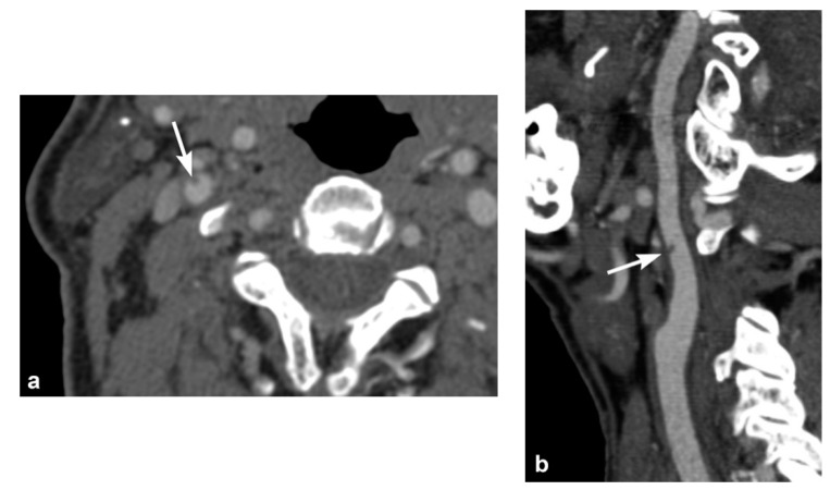
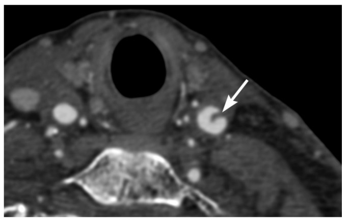
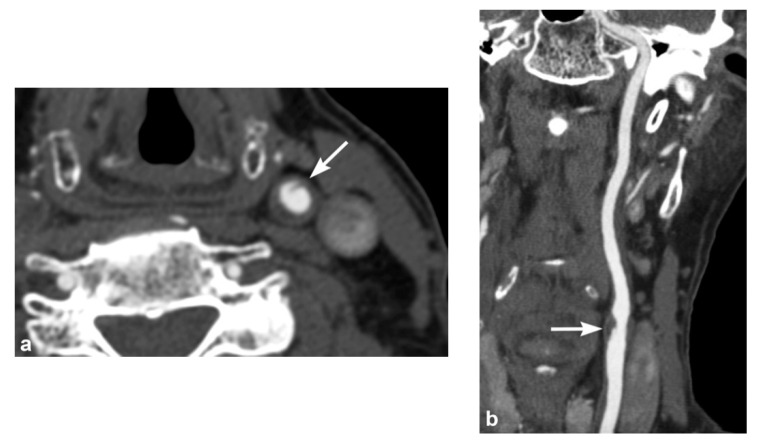
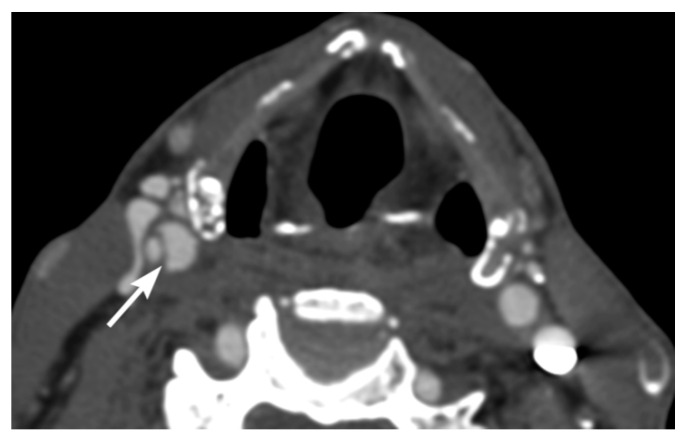
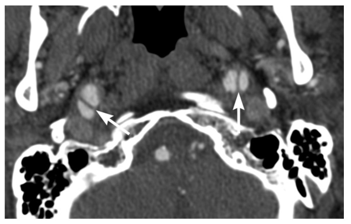
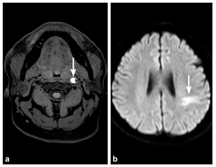
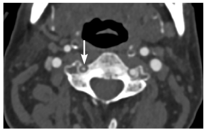
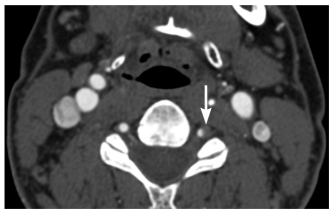
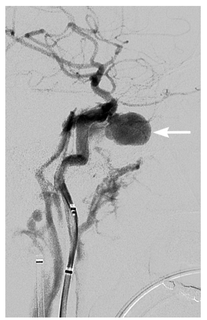
Blunt cerebrovascular injury (BCVI) is an often underrecognized injury occurring in the carotid or vertebral arteries, associated with a risk of ischemic stroke and potential for poor neurological outcome or death. Computed tomographic angiography (CTA) is the most common modality for initial screening and diagnosis. Vessel wall intimal injuries, intraluminal thrombus, dissection, intramural hematoma, pseudoaneurysm, vessel transection, and arteriovenous fistula, are potential findings to be considered in approach to imaging. Identification of high-risk trauma patients based on clinical and radiological risk factors can determine patients at risk of BCVI for targeted screening.
Keywords: blunt cerebrovascular injury; carotid arteries; neck injuries; stroke; vertebral arteries.
Conflict of interest statement
The authors declare no conflict of interest.
Figures
Similar articles
-
Association between cervical fracture patterns and blunt cerebrovascular injury when screened with computed tomographic angiography.Spine J. 2024 Feb;24(2):310-316. doi: 10.1016/j.spinee.2023.09.010. Epub 2023 Sep 20. Spine J. 2024. PMID: 37734494
-
Risk factors for traumatic blunt cerebrovascular injury diagnosed by computed tomography angiography in the pediatric population: a retrospective cohort study.J Neurosurg Pediatr. 2015 Jun;15(6):599-606. doi: 10.3171/2014.11.PEDS14397. Epub 2015 Mar 6. J Neurosurg Pediatr. 2015. PMID: 25745952
-
Blunt cerebrovascular injuries: imaging with multidetector CT angiography.Radiographics. 2008 Oct;28(6):1689-708; discussion 1709-10. doi: 10.1148/rg.286085521. Radiographics. 2008. PMID: 18936030 Review.
-
Initial screening test for blunt cerebrovascular injury: Validity assessment of whole-body computed tomography.Surgery. 2015 Sep;158(3):627-35. doi: 10.1016/j.surg.2015.03.063. Epub 2015 Jun 9. Surgery. 2015. PMID: 26067461
-
Imaging and Management of Blunt Cerebrovascular Injury.Radiographics. 2018 Mar-Apr;38(2):542-563. doi: 10.1148/rg.2018170140. Radiographics. 2018. PMID: 29528828 Free PMC article. Review.
Cited by
-
Shifting Perspectives: Equal Blunt Cerebrovascular Risk in Low-Versus High-Energy Cervical Fracture.J Surg Res. 2024 Aug;300:63-70. doi: 10.1016/j.jss.2024.04.058. Epub 2024 May 24. J Surg Res. 2024. PMID: 38795674 Free PMC article.
-
Emergent neurovascular imaging in patients with blunt traumatic injuries.Front Radiol. 2022 Sep 15;2:1001114. doi: 10.3389/fradi.2022.1001114. eCollection 2022. Front Radiol. 2022. PMID: 37492683 Free PMC article. Review.
-
Experience and lessons learned from blunt cerebrovascular injuries: A case report.Heliyon. 2023 Feb 20;9(3):e13968. doi: 10.1016/j.heliyon.2023.e13968. eCollection 2023 Mar. Heliyon. 2023. PMID: 36895340 Free PMC article.
-
Imaging of cerebrovascular complications from blunt skull base trauma.Emerg Radiol. 2024 Aug;31(4):529-542. doi: 10.1007/s10140-024-02243-z. Epub 2024 May 28. Emerg Radiol. 2024. PMID: 38806851 Free PMC article. Review.
-
The effect of circle of willis anatomy and scanning practices on outcomes for blunt cerebrovascular injuries.Scand J Trauma Resusc Emerg Med. 2024 Jun 17;32(1):57. doi: 10.1186/s13049-024-01225-x. Scand J Trauma Resusc Emerg Med. 2024. PMID: 38886775 Free PMC article.
References
-
- Berne J.D., Reuland K.S., Villarreal D.H., McGovern T.M., Rowe S.A., Norwood S.H. Sixteen-Slice Multi-Detector Computed Tomographic angiography improves the accuracy of screening for blunt cerebrovascular injury. J. Trauma Acute Care Surg. 2006;60:1204–1210. doi: 10.1097/01.ta.0000220435.55791.ce. - DOI - PubMed
Publication types
MeSH terms
LinkOut - more resources
Full Text Sources
