

IRIS-Intelligent Rapid Interactive Segmentation for Measuring Liver Cyst Volumes in Autosomal Dominant Polycystic Kidney Disease
- PMID: 35202202
- PMCID: PMC8877996
- DOI: 10.3390/tomography8010037
IRIS-Intelligent Rapid Interactive Segmentation for Measuring Liver Cyst Volumes in Autosomal Dominant Polycystic Kidney Disease
Abstract
Purpose: To develop and integrate interactive features with automatic methods for accurate liver cyst segmentation in patients with autosomal dominant polycystic kidney and liver disease (ADPKD).
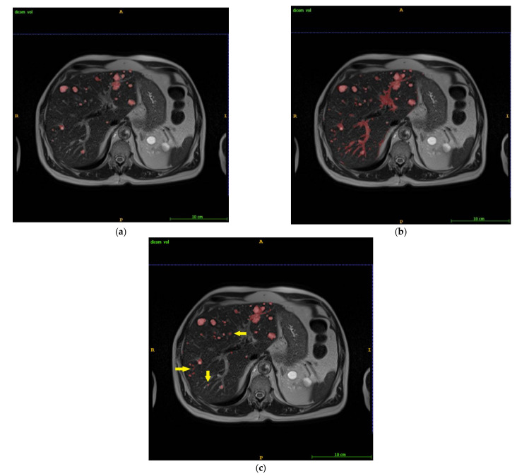
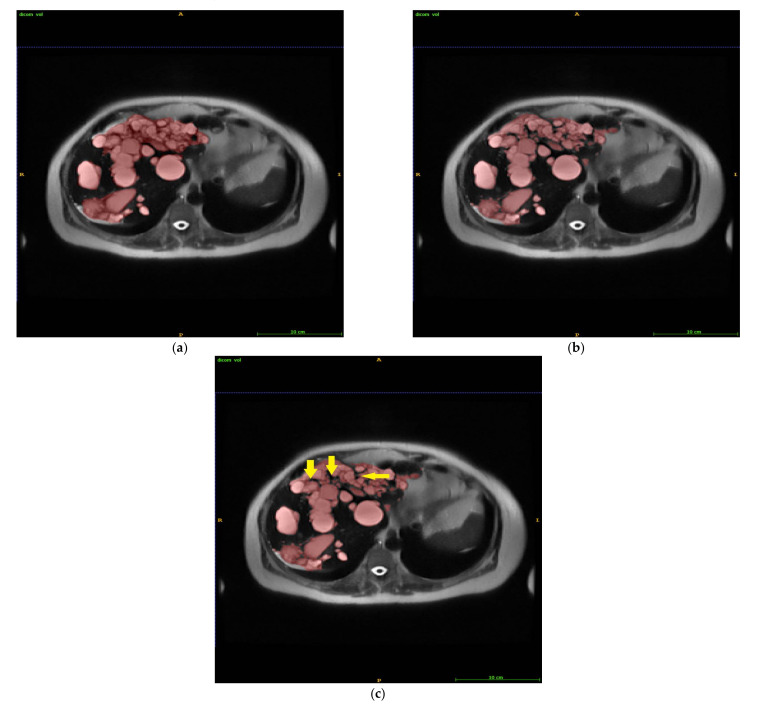
Methods: SmartClick and antiSmartClick were developed using iterative region growth guided by spatial and intensity connections and were integrated with automated level set (LS) segmentation and graphical user interface, forming an intelligent rapid interactive segmentation (IRIS) tool. IRIS and LS segmentations of liver cysts on T2 weighted images of patients with ADPKD (n = 17) were compared with manual segmentation as ground truth (GT).
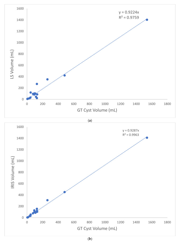
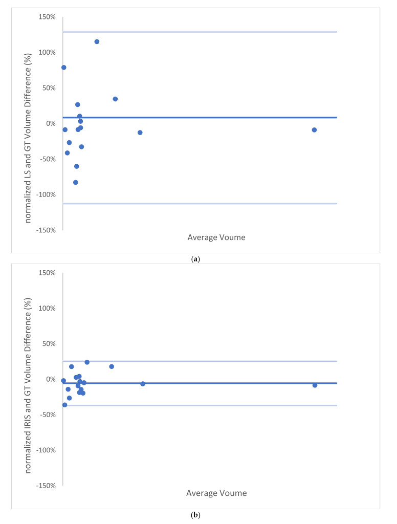
Results: Compared to manual GT, IRIS reduced the segmentation time by more than 10-fold. Compared to automated LS, IRIS reduced the mean liver cyst volume error from 42.22% to 13.44% (p < 0.001). IRIS segmentation agreed well with manual GT (79% dice score and 99% intraclass correlation coefficient).
Conclusion: IRIS is feasible for fast, accurate liver cyst segmentation in patients with ADPKD.
Keywords: intelligent rapid interactive segmentation; lesion segmentation; liver cyst.
Conflict of interest statement
The authors declare no conflict of interest.
Figures
References
-
- Van Keimpema L., De Koning D.B., Van Hoek B., Van Den Berg A.P., Van Oijen M.G., De Man R.A., Nevens F., Drenth J.P. Patients with isolated polycystic liver disease referred to liver centres: Clinical characterization of 137 cases. Liver Int. 2011;31:92–98. doi: 10.1111/j.1478-3231.2010.02247.x. - DOI - PubMed
-
- Malmberg F., Nordenskjold R., Strand R., Kullberg J. SmartPaint: A tool for interactive segmentation of medical volume images. Comput. Methods Biomech. Biomed. Eng.-Imaging Vis. 2017;5:36–44. doi: 10.1080/21681163.2014.960535. - DOI
-
- Kim Y., Bae S.K., Cheng T., Tao C., Ge Y., Chapman A.B., Torres V.E., Yu A.S.L., Mrug M., Bennett W.M., et al. Automated segmentation of liver and liver cysts from bounded abdominal MR images in patients with autosomal dominant polycystic kidney disease. Phys. Med. Biol. 2016;61:7864–7880. doi: 10.1088/0031-9155/61/22/7864. - DOI - PMC - PubMed
Publication types
MeSH terms
Grants and funding
LinkOut - more resources
Full Text Sources
Medical
Miscellaneous
