

Integrating the OHIF Viewer into XNAT: Achievements, Challenges and Prospects for Quantitative Imaging Studies
- PMID: 35202205
- PMCID: PMC8875191
- DOI: 10.3390/tomography8010040
Integrating the OHIF Viewer into XNAT: Achievements, Challenges and Prospects for Quantitative Imaging Studies
Abstract
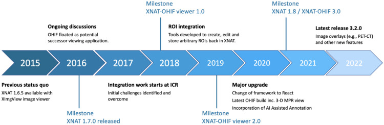
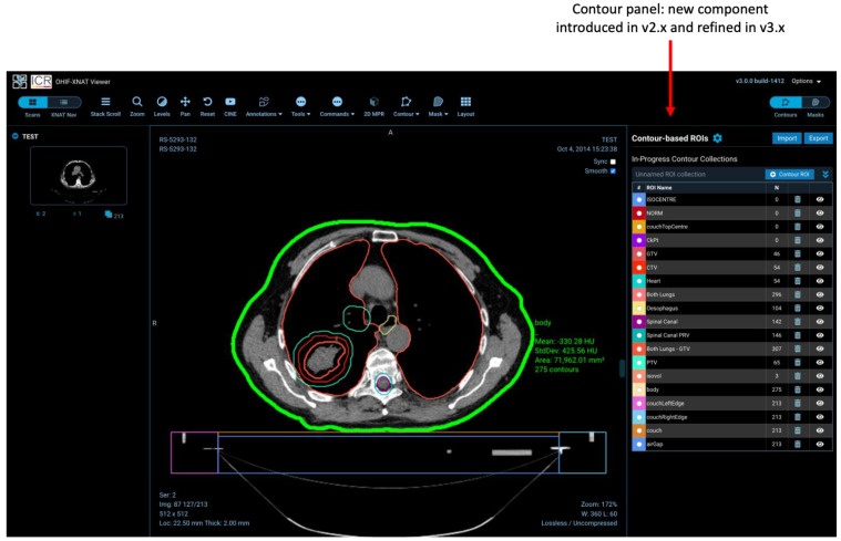
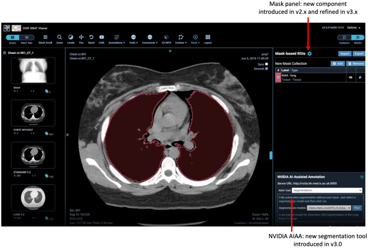
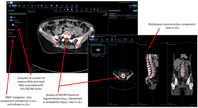
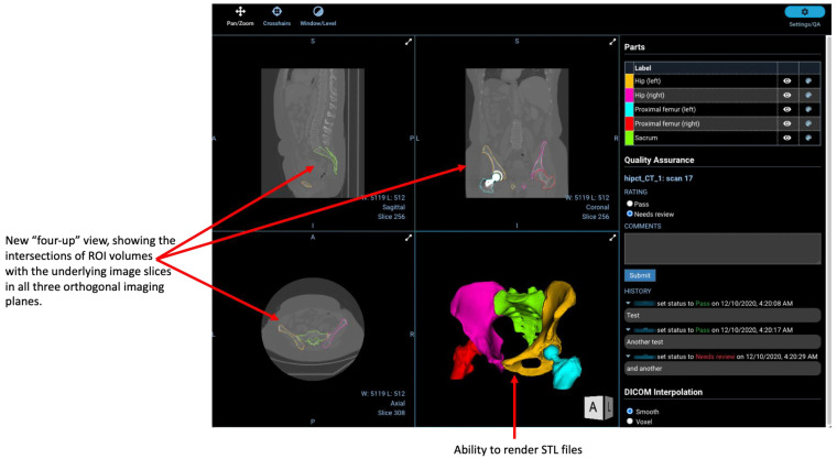
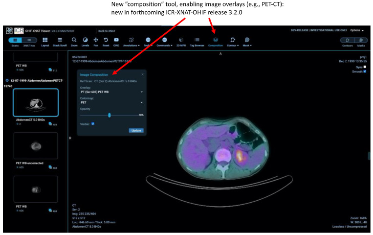
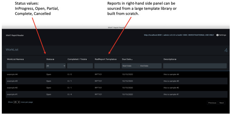
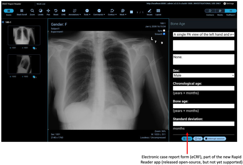
Purpose: XNAT is an informatics software platform to support imaging research, particularly in the context of large, multicentre studies of the type that are essential to validate quantitative imaging biomarkers. XNAT provides import, archiving, processing and secure distribution facilities for image and related study data. Until recently, however, modern data visualisation and annotation tools were lacking on the XNAT platform. We describe the background to, and implementation of, an integration of the Open Health Imaging Foundation (OHIF) Viewer into the XNAT environment. We explain the challenges overcome and discuss future prospects for quantitative imaging studies. Materials and methods: The OHIF Viewer adopts an approach based on the DICOM web protocol. To allow operation in an XNAT environment, a data-routing methodology was developed to overcome the mismatch between the DICOM and XNAT information models and a custom viewer panel created to allow navigation within the viewer between different XNAT projects, subjects and imaging sessions. Modifications to the development environment were made to allow developers to test new code more easily against a live XNAT instance. Major new developments focused on the creation and storage of regions-of-interest (ROIs) and included: ROI creation and editing tools for both contour- and mask-based regions; a "smart CT" paintbrush tool; the integration of NVIDIA's Artificial Intelligence Assisted Annotation (AIAA); the ability to view surface meshes, fractional segmentation maps and image overlays; and a rapid image reader tool aimed at radiologists. We have incorporated the OHIF microscopy extension and, in parallel, introduced support for microscopy session types within XNAT for the first time. Results: Integration of the OHIF Viewer within XNAT has been highly successful and numerous additional and enhanced tools have been created in a programme started in 2017 that is still ongoing. The software has been downloaded more than 3700 times during the course of the development work reported here, demonstrating the impact of the work. Conclusions: The OHIF open-source, zero-footprint web viewer has been incorporated into the XNAT platform and is now used at many institutions worldwide. Further innovations are envisaged in the near future.
Keywords: OHIF; XNAT; image visualisation; rapid reader; regions-of-interest; web viewer.
Conflict of interest statement
The funders had no role in the design of the study; in the collection, analyses, or interpretation of data; in the writing of the manuscript, or in the decision to publish the results. E.O.A. is a member of the Editorial Board of Tomography. G.J.H. is a member of Novometrics LLC and IQ Medical Imaging LLC and an advisor for Fovia Inc. E.S. is co-founder and shareholder of Lucida Medical Ltd. L.E.S. has received consulting fees from Lucida Medical Ltd.
Figures
References
-
- Ziegler E., Urban T., Brown D., Petts J., Pieper S.D., Lewis R., Hafey C., Harris G.J. Open Health Imaging Foundation Viewer: An Extensible Open-Source Framework for Building Web-Based Imaging Applications to Support Cancer Research. JCO Clin. Cancer Inform. 2020;4:336–345. doi: 10.1200/CCI.19.00131. - DOI - PMC - PubMed
-
- The Open Health Imaging Foundation. [(accessed on 14 July 2021)]. Available online: www.ohif.org.
-
- Fielding R.T., Taylor R.N. Principled design of the modern Web architecture. ACM Trans. Internet Technol. 2002;2:115–150. doi: 10.1145/514183.514185. - DOI
Publication types
MeSH terms
Grants and funding
LinkOut - more resources
Full Text Sources
Medical
