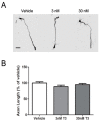
Triiodothyronine or Antioxidants Block the Inhibitory Effects of BDE-47 and BDE-49 on Axonal Growth in Rat Hippocampal Neuron-Glia Co-Cultures
- PMID: 35202279
- PMCID: PMC8879960
- DOI: 10.3390/toxics10020092
Triiodothyronine or Antioxidants Block the Inhibitory Effects of BDE-47 and BDE-49 on Axonal Growth in Rat Hippocampal Neuron-Glia Co-Cultures
Abstract
We previously demonstrated that polybrominated diphenyl ethers (PBDEs) inhibit the growth of axons in primary rat hippocampal neurons. Here, we test the hypothesis that PBDE effects on axonal morphogenesis are mediated by thyroid hormone and/or reactive oxygen species (ROS)-dependent mechanisms. Axonal growth and ROS were quantified in primary neuronal-glial co-cultures dissociated from neonatal rat hippocampi exposed to nM concentrations of BDE-47 or BDE-49 in the absence or presence of triiodothyronine (T3; 3-30 nM), N-acetyl-cysteine (NAC; 100 µM), or α-tocopherol (100 µM). Co-exposure to T3 or either antioxidant prevented inhibition of axonal growth in hippocampal cultures exposed to BDE-47 or BDE-49. T3 supplementation in cultures not exposed to PBDEs did not alter axonal growth. T3 did, however, prevent PBDE-induced ROS generation and alterations in mitochondrial metabolism. Collectively, our data indicate that PBDEs inhibit axonal growth via ROS-dependent mechanisms, and that T3 protects axonal growth by inhibiting PBDE-induced ROS. These observations suggest that co-exposure to endocrine disruptors that decrease TH signaling in the brain may increase vulnerability to the adverse effects of developmental PBDE exposure on axonal morphogenesis.
Keywords: PBDE; axonal growth; developmental neurotoxicity; neuronal morphogenesis; reactive oxygen species; thyroid hormone.
Conflict of interest statement
The authors declare no conflict of interest. The funders had no role in the design of the study; in the collection, analyses, or interpretation of data; in the writing of the manuscript, or in the decision to publish the results.
Figures


Similar articles
-
Distribution of polybrominated diphenyl ethers (PBDEs) in placental tissues of maternal and fetal origin in exposed Wistar rats and associations with thyroid hormone levels.Toxicol Sci. 2025 Mar 1;204(1):20-30. doi: 10.1093/toxsci/kfae151. Toxicol Sci. 2025. PMID: 39626304
-
Polybrominated diphenyl ethers (PBDEs) in household dust: A systematic review on spatio-temporal distribution, sources, and health risk assessment.Chemosphere. 2023 Feb;314:137641. doi: 10.1016/j.chemosphere.2022.137641. Epub 2022 Dec 27. Chemosphere. 2023. PMID: 36584828
-
From the Cover: BDE-47 and BDE-49 Inhibit Axonal Growth in Primary Rat Hippocampal Neuron-Glia Co-Cultures via Ryanodine Receptor-Dependent Mechanisms.Toxicol Sci. 2017 Apr 1;156(2):375-386. doi: 10.1093/toxsci/kfw259. Toxicol Sci. 2017. PMID: 28003438 Free PMC article.
-
Socioeconomic and racial-ethnic disparities in flame retardant exposure and executive function skills in preschool children.Environ Health. 2025 Jul 8;24(1):46. doi: 10.1186/s12940-025-01200-8. Environ Health. 2025. PMID: 40629376 Free PMC article.
-
Associations between human exposure to polybrominated diphenyl ether flame retardants via diet and indoor dust, and internal dose: A systematic review.Environ Int. 2016 Jul-Aug;92-93:680-94. doi: 10.1016/j.envint.2016.02.017. Epub 2016 Apr 9. Environ Int. 2016. PMID: 27066981
Cited by
-
Effect of the exposure to brominated flame retardants on hyperuricemia using interpretable machine learning algorithms based on the SHAP methodology.PLoS One. 2025 Jun 26;20(6):e0325896. doi: 10.1371/journal.pone.0325896. eCollection 2025. PLoS One. 2025. PMID: 40570004 Free PMC article.
References
-
- Lam J., Lanphear B.P., Bellinger D., Axelrad D.A., McPartland J., Sutton P., Davidson L., Daniels N., Sen S., Woodruff T.J. Developmental PBDE Exposure and IQ/ADHD in Childhood: A Systematic Review and Meta-analysis. Environ. Health Perspect. 2017;125:086001. doi: 10.1289/EHP1632. - DOI - PMC - PubMed
-
- Vuong A.M., Braun J.M., Webster G.M., Thomas Zoeller R., Hoofnagle A.N., Sjödin A., Yolton K., Lanphear B.P., Chen A. Polybrominated Diphenyl Ether (PBDE) Exposures and Thyroid Hormones in Children at Age 3 Years. Environ. Int. 2018;117:339–347. doi: 10.1016/j.envint.2018.05.019. - DOI - PMC - PubMed
-
- Dorman D.C., Chiu W., Hales B.F., Hauser R., Johnson K.J., Mantus E., Martel S., Robinson K.A., Rooney A.A., Rudel R., et al. Polybrominated Diphenyl Ether (PBDE) Neurotoxicity: A Systematic Review and Meta-Analysis of Animal Evidence. J. Toxicol. Environ. Health B Crit. Rev. 2018;21:269–289. doi: 10.1080/10937404.2018.1514829. - DOI - PMC - PubMed

