

Auricular Non-Epithelial Tumors with Solar Elastosis in Cats: A Possible UV-Induced Pathogenesis
- PMID: 35202288
- PMCID: PMC8874616
- DOI: 10.3390/vetsci9020034
Auricular Non-Epithelial Tumors with Solar Elastosis in Cats: A Possible UV-Induced Pathogenesis
Abstract
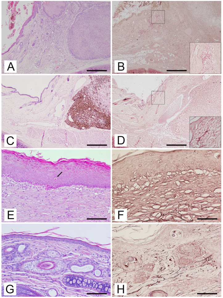
The photoinduced etiopathology of actinic keratosis and squamous cell carcinoma in feline species is well known. This etiology has also been reported for non-epithelial cutaneous tumors in other species. To date, no cases of auricular non-epithelial cutaneous neoplasms erased in a contest of actinic keratosis in cats have been reported. The aim of this study was to describe feline auricular non-epithelial cutaneous neoplasms associated with typical UV-induced cutaneous lesions and solar elastosis. The study was conducted on five feline cases diagnosed with auricular non-epithelial cutaneous tumors (two fibrosarcomas, one mixosarcoma, one epithelioid melanoma and one hemangiosarcoma), selected from the Tumor Registry of the Department of Veterinary Sciences of the University of Pisa (1998-2018). Ten and six feline auricular biopsies of normal skin and skin with actinic keratosis, respectively, were used as controls. Orcein stain was used to investigate solar elastosis. Histological changes related to chronic solar irradiation were documented in the skin adjacent to the neoplastic lesions in the five cats. Considering the anatomical localization and the results of histopathology, this study suggests that non-epithelial cutaneous neoplasms may have a UV-induced etiopathogenesis in the feline species.
Keywords: UV light; auricular; cats; elastosis; neoplasms; solar.
Conflict of interest statement
The authors declare no conflict of interest.
Figures
Similar articles
-
Skin Photodamage Lesions in a Bilateral Feline Auricular Primary Fibrosarcoma.Vet Sci. 2022 Oct 5;9(10):548. doi: 10.3390/vetsci9100548. Vet Sci. 2022. PMID: 36288161 Free PMC article.
-
Solar elastosis and cutaneous melanoma: a site-specific analysis.Int J Cancer. 2015 Jun 15;136(12):2900-11. doi: 10.1002/ijc.29335. Epub 2014 Nov 26. Int J Cancer. 2015. PMID: 25403328
-
Degree of Actinic Elastosis Is a Surrogate of Exposure to Chronic Ultraviolet Radiation and Correlates More Strongly with Cutaneous Squamous Cell Carcinoma than Basal Cell Carcinoma.Life (Basel). 2023 Mar 17;13(3):811. doi: 10.3390/life13030811. Life (Basel). 2023. PMID: 36983966 Free PMC article.
-
Pathology and pathobiology of actinic (solar) keratosis - an update.Br J Dermatol. 2007 Dec;157 Suppl 2:18-20. doi: 10.1111/j.1365-2133.2007.08267.x. Br J Dermatol. 2007. PMID: 18067626 Review.
-
[Actinic keratosis, Bowen's disease, keratoacanthoma and squamous cell carcinoma of the skin].Pathologe. 2015 Feb;36(1):16-29. doi: 10.1007/s00292-014-2063-3. Pathologe. 2015. PMID: 25663185 Review. German.
Cited by
-
Skin Cancer Metabolic Profile Assessed by Different Analytical Platforms.Int J Mol Sci. 2023 Jan 13;24(2):1604. doi: 10.3390/ijms24021604. Int J Mol Sci. 2023. PMID: 36675128 Free PMC article. Review.
-
Skin Photodamage Lesions in a Bilateral Feline Auricular Primary Fibrosarcoma.Vet Sci. 2022 Oct 5;9(10):548. doi: 10.3390/vetsci9100548. Vet Sci. 2022. PMID: 36288161 Free PMC article.
-
Electroporation in Translational Medicine: From Veterinary Experience to Human Oncology.Cancers (Basel). 2024 Mar 6;16(5):1067. doi: 10.3390/cancers16051067. Cancers (Basel). 2024. PMID: 38473422 Free PMC article. Review.
References
-
- Dunstan R.W., Credille K.M., Walder E.J. The light and the skin. In: Kwochka K.W., Willemse T., von Tscharner C., editors. Advances in Veterinary Dermatology. Butterworth Heinemann; Oxford, UK: 1998. pp. 3–35.
LinkOut - more resources
Full Text Sources
Miscellaneous
