Review
. 2022 Jan 26;9(2):46.
doi: 10.3390/vetsci9020046.

Feline Uveal Melanoma Review: Our Current Understanding and Recent Research Advances

Affiliations
Review

Feline Uveal Melanoma Review: Our Current Understanding and Recent Research Advances

David Kayes et al. Vet Sci. .

Abstract

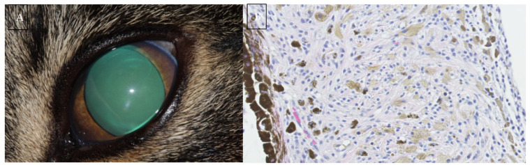
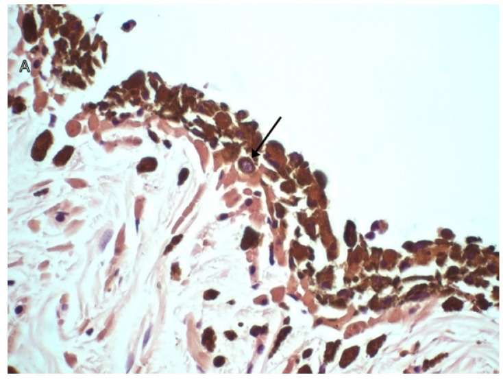
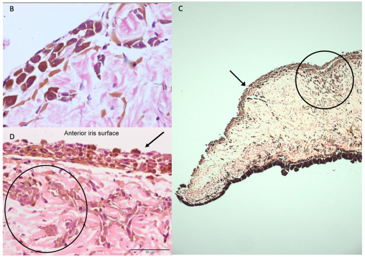
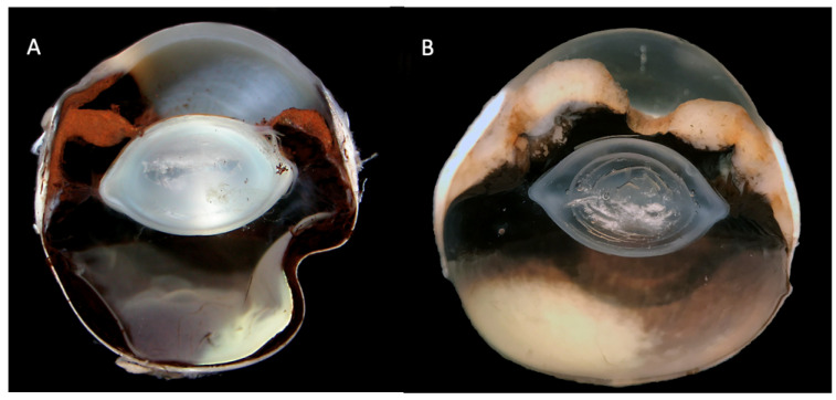
Melanocytic neoplasia is the most common form of ocular tumour in cats, accounting for 67% of cases in an analysis of 2614 cases of primary ocular neoplasia. Feline diffuse iris melanoma (FDIM) is by far the most common form of ocular melanocytic neoplasia, with limbal melanomas and atypical melanoma (melanoma affecting the choroid or ciliary body) infrequently recognised. Early lesions begin as flat areas of pigmentation of the iris, known as iris melanosis. This melanosis is a precursor lesion that can become FDIM when pigmented cells infiltrate the anterior iris stroma, commonly alongside a transition in cell morphology. The differentiation between FDIM and benign iris melanosis is only recognisable though histologic examination, with no in vivo means of identifying the malignant transformation. The behaviour of FDIM is variable and difficult to predict. Some FDIM lesions have a more benign progression and can slowly grow or remain static for years without affecting the ocular or systemic health of the individual, whilst other tumours behave aggressively, invading the ocular structures and significantly affecting the life expectancy of cats through metastatic disease. This makes management and timely enucleation of these cases challenging in practice. This article aims to review our current knowledge of FDIM.

Keywords: feline; iris; melanoma; melanosis; uveal.


Conflict of interest statement

The authors declare no conflict of interest.

Figures

Figure 1
Figure 1
(A) Multifocal iris hyperpigmentation in a 4-year-old Domestic Shorthair. (B) Extensive, diffuse hyperpigmentation of the iris of a 7-year-old Domestic Shorthair with appearance of the iridocorneal drainage angle in the same eye (C). Gonioscopy is performed routinely for all cases presenting with a suspicion of feline diffuse iris melanoma (FDIM). The hyperpigmentation in both cases may represent iris melanosis or FDIM and iris biopsy should be considered.
Figure 2
Figure 2
(A) Photograph of a 12-year-old Domestic Shorthair presenting for unilateral glaucoma. Subsequent enucleation resulted in a histopathologic diagnosis of feline diffuse iris melanoma. (B) A high magnification (400×) H&E section from the same case. Note the heavily pigmented posterior iris epithelium aiding orientation of the image. The iris is infiltrated with spindle to round cells. Most cells have a lightly pigmented brown granular cytoplasm with scattered highly pigmented cells. Nuclei are round to ovoid with coarsely stippled chromatin and often a single prominent nucleolus. Anisocytosis and anisokaryosis are mild and mitoses are rare which is not typical of FDIM. Image (B) courtesy of Linda Morrison and Alexandra Malbon (University of Edinburgh Pathology Department).
Figure 3
Figure 3
H&E histopathology sections of the feline iris. (A) A high magnification (×600) section showing a melanocyte with a prominent, open nucleus with a small distinct nucleolus, indicating nuclear atypia. (B) A high magnification (×600) section of an early case of FDIM with the very start of invasion of the stroma with dysplastic melanocytes. A low magnification (×100) (C) and a high magnification (×400) (D) section of two separate cases of FDIM. Dysplastic melanocytes with anisokariosis and nuclear atypia line the anterior iris stroma (arrows) and invade the iris stroma (circles). Images courtesy of Emma J. Scurrell (CytoPath Veterinary Pathology).
Figure 3
Figure 3
H&E histopathology sections of the feline iris. (A) A high magnification (×600) section showing a melanocyte with a prominent, open nucleus with a small distinct nucleolus, indicating nuclear atypia. (B) A high magnification (×600) section of an early case of FDIM with the very start of invasion of the stroma with dysplastic melanocytes. A low magnification (×100) (C) and a high magnification (×400) (D) section of two separate cases of FDIM. Dysplastic melanocytes with anisokariosis and nuclear atypia line the anterior iris stroma (arrows) and invade the iris stroma (circles). Images courtesy of Emma J. Scurrell (CytoPath Veterinary Pathology).
Figure 4
Figure 4
Gross section of a feline globe with diffuse iris melanoma (FDIM) (A) and an amelanotic FDIM variant (B). Note the marked iris thickening in both sections due to infiltration with neoplastic melanocytes. Images courtesy of Emma J. Scurrell (Cytopath Veterinary Pathology).

References

    1. Dubielzig R.R. World Small Animal Veterinary Association World Congress Proceedings. VIN.com. 2011. [(accessed on 29 May 2021)]. Available online: https://www.vin.com/apputil/content/defaultadv1.aspx?pId=11343&meta=VIN&....
    1. Duncan D.E., Peiffer R.L. Morphology and prognostic indicators of anterior uveal melanomas in cats. Prog. Vet. Comp. Ophthalmol. 1991;1:25–32.
    1. Dubielzig R.R., Lindley D.M. The relationship between pigmented spots on the feline iris and diffuse iris melanoma (abstract 96) Vet. Pathol. 1993;30:451.
    1. Gelatt K.N., Gilger B.C., Kern T.J. Veterinary Ophthalmology. Volume 28. Wiley-Blackwell; Hoboken, NJ, USA: 2021. p. 1715.
    1. Kalishman J.B., Chappell R., Flood L.A., Dubielzig R.R. A matched observational study of survival in cats with enucleation due to diffuse iris melanoma. Vet. Ophthalmol. 1998;1:25–29. doi: 10.1046/j.1463-5224.1998.00006.x. - DOI - PubMed

LinkOut - more resources