

In Situ Hybridization of Feline Leukemia Virus in a Case of Osteochondromatosis
- PMID: 35202311
- PMCID: PMC8878754
- DOI: 10.3390/vetsci9020059
In Situ Hybridization of Feline Leukemia Virus in a Case of Osteochondromatosis
Abstract
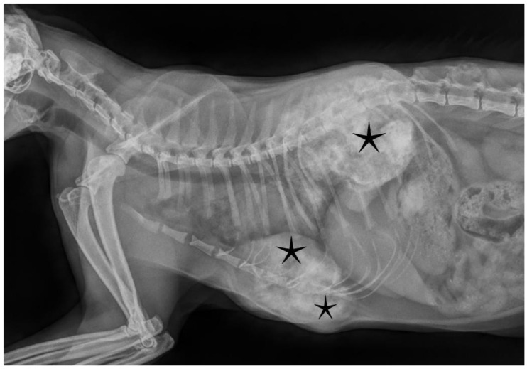
Osteochondromatosis, also known as multiple cartilaginous exostosis, polyostotic osteochondroma, and multiple osteochondromas, comprises one-fifth of all primary bone tumors in cats, with no breed or sex predisposition or hereditary pattern. Unlike in dogs, horses, and humans, it is predominantly seen in young cats (2-4 years old), after the maturation of the skeleton. Although the pathogenesis of osteochondromatosis is not fully understood, it is considered to be related to infection by feline leukemia virus (FeLV) or other retroviruses, such as the feline sarcoma virus. However, the presence of viral particles within tumor lesions has only been demonstrated by electron microscopy. The malignant transformation of osteochondromas, most typically to osteosarcoma or chondrosarcoma, has also been attributed to the viral infection. Here we report the case of osteochondromatosis in a 3.5-year-old male domestic European shorthair cat with concurrent FeLV infection confirmed by polymerase chain reaction. Viral RNA was visualized in representative tissues (spleen, mesenteric lymph node, liver, kidney, lung, brain) and in the osteochondromas with RNAscope in situ hybridization, which supports that FeLV infection may be involved in the pathogenesis of osteochondromatosis.
Keywords: FeLV; feline; in situ hybridization; osteochondromatosis.
Conflict of interest statement
The authors declare no conflict of interest.
Figures


Similar articles
-
Feline osteochondromatosis in a 12-year-old feline leukaemia virus-negative cat.J Comp Pathol. 2023 Aug;205:24-26. doi: 10.1016/j.jcpa.2023.07.003. Epub 2023 Aug 17. J Comp Pathol. 2023. PMID: 37597496
-
Feline osteochondromatosis in a FELV-negative European shorthair cat.Tierarztl Prax Ausg K Kleintiere Heimtiere. 2014;42(1):55-9. Tierarztl Prax Ausg K Kleintiere Heimtiere. 2014. PMID: 24518948
-
A frameshift variant in the EXT1 gene in a feline leukemia virus-negative cat with osteochondromatosis.Anim Genet. 2022 Oct;53(5):696-699. doi: 10.1111/age.13232. Epub 2022 Jun 20. Anim Genet. 2022. PMID: 35719100
-
Quantification of endogenous and exogenous feline leukemia virus sequences by real-time PCR assays.Vet Immunol Immunopathol. 2008 May 15;123(1-2):129-33. doi: 10.1016/j.vetimm.2008.01.027. Epub 2008 Jan 19. Vet Immunol Immunopathol. 2008. PMID: 18295344 Review.
-
Feline leukemia virus infection and diseases.J Am Vet Med Assoc. 1991 Nov 15;199(10):1287-97. J Am Vet Med Assoc. 1991. PMID: 1666070 Review.
References
-
- Thompson K.G., Pool R.R. Tumors of bones. In: Meuten D.J., editor. Tumors in Domestic Animals. 5th ed. John Wiley & Sons, Inc.; Ames, IA, USA: 2017. pp. 368–369.
-
- de Oliveira Reis M., Santos de Mello L., Hesse K.L., Lorenzett M.P., dos Reis K.D.H.L., Campos F.S., Roehe P.M., Pavarin S.P. Osteochondroma in a young cat infected by feline leukemia virus. Ciênc. Rural. 2017;47:1. doi: 10.1590/0103-8478cr20151558. - DOI
-
- Nolff M.C., Puff C., Länger B., Fehr M. Feline osteochondromatosis in a FeLV-negative European shorthair cat. Tierärztliche Prax. Kleintiere. 2014;42:55–59. - PubMed
Publication types
LinkOut - more resources
Full Text Sources
Miscellaneous
