. 2022 Feb 24;18(2):e1010084.
doi: 10.1371/journal.pgen.1010084. eCollection 2022 Feb.

FXS causing missense mutations disrupt FMRP granule formation, dynamics, and function

Affiliations

FXS causing missense mutations disrupt FMRP granule formation, dynamics, and function

Emily L Starke et al. PLoS Genet. .

Abstract

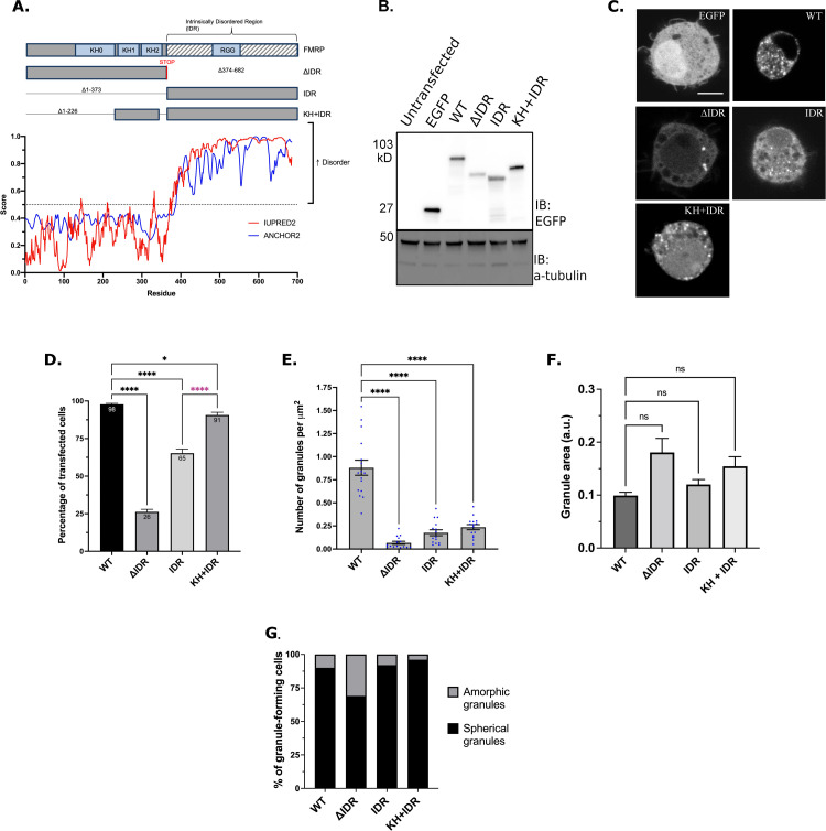
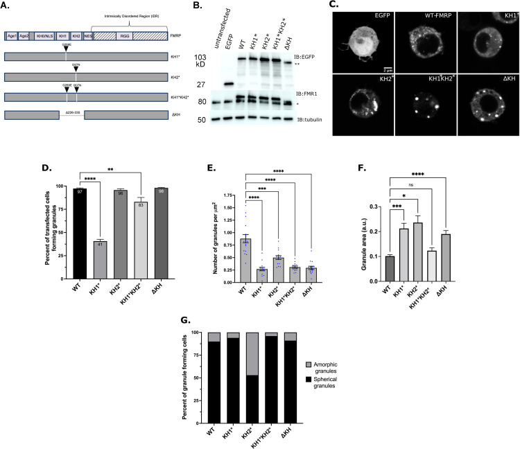
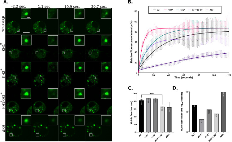
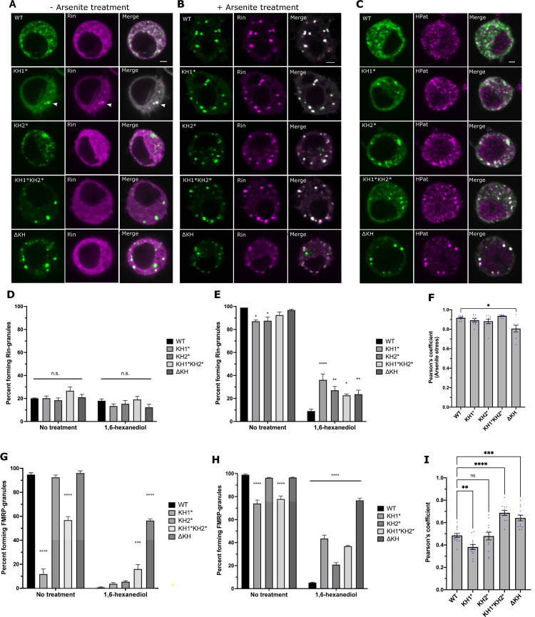
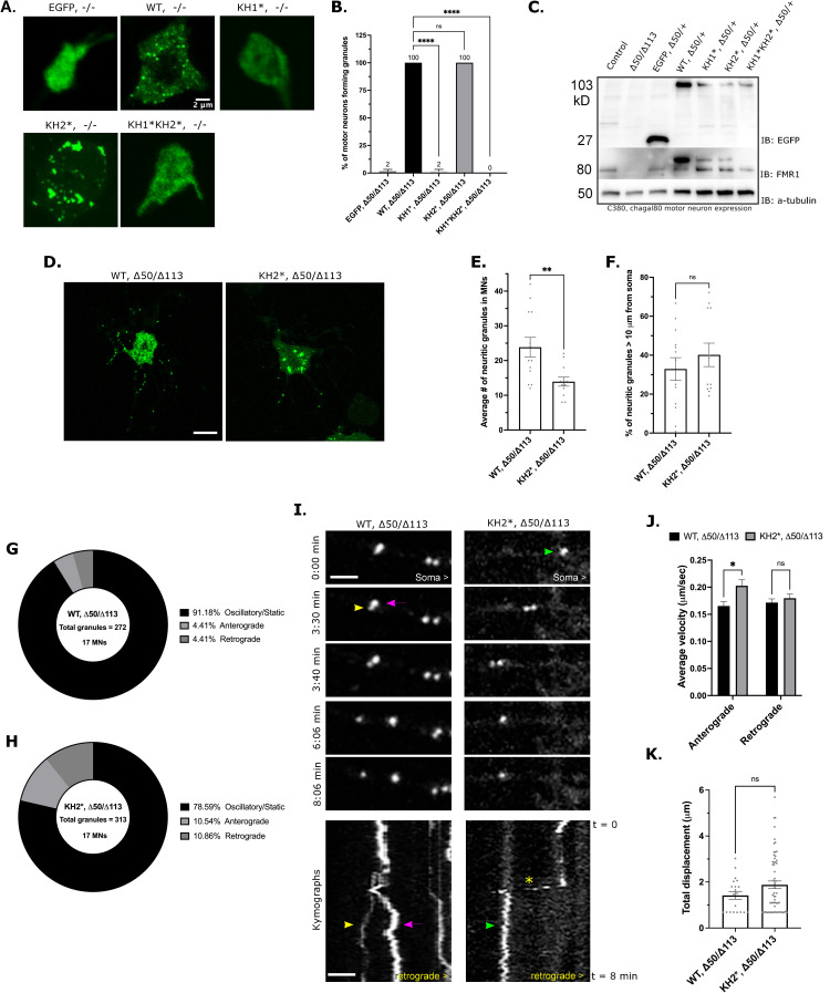
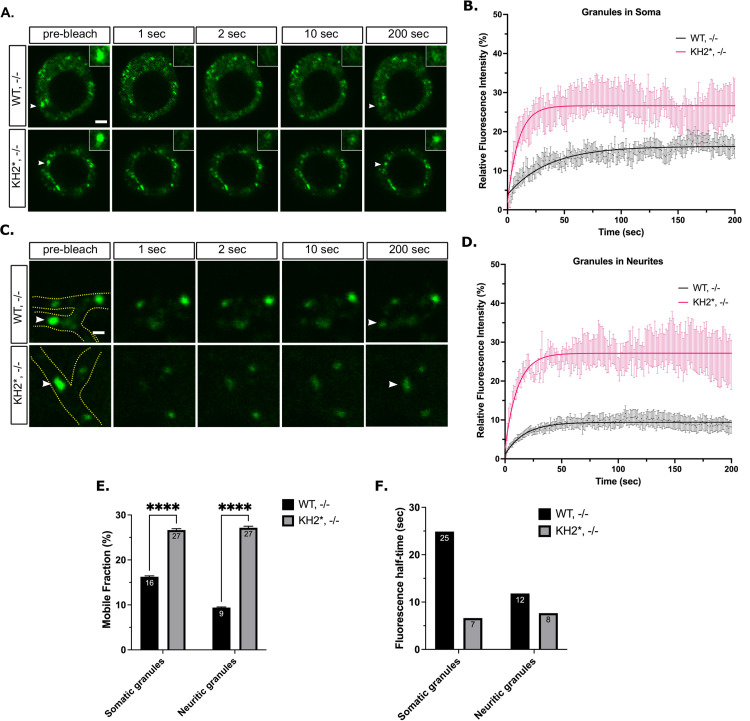
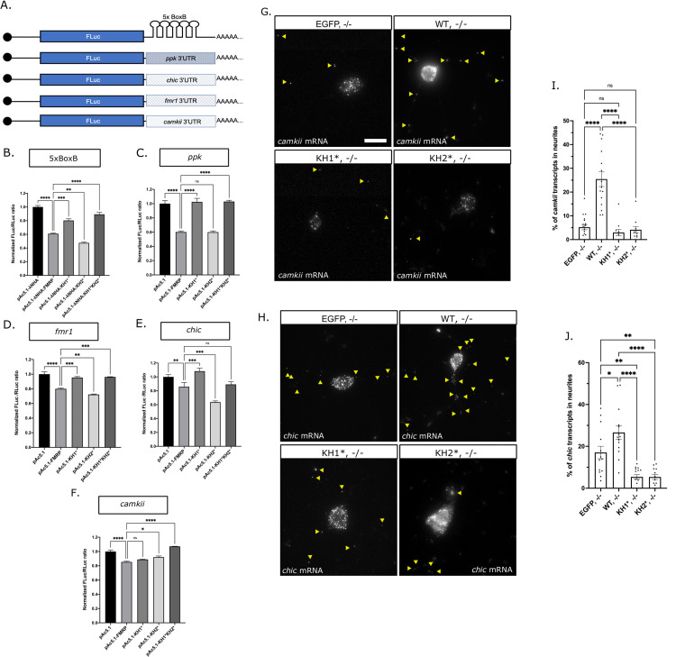
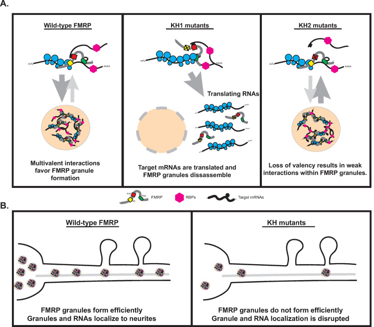
Fragile X Syndrome (FXS) is the most prevalent cause of inherited mental deficiency and is the most common monogenetic cause of autism spectral disorder (ASD). Here, we demonstrate that disease-causing missense mutations in the conserved K homology (KH) RNA binding domains (RBDs) of FMRP cause defects in its ability to form RNA transport granules in neurons. Using molecular, genetic, and imaging approaches in the Drosophila FXS model system, we show that the KH1 and KH2 domains of FMRP regulate distinct aspects of neuronal FMRP granule formation, dynamics, and transport. Furthermore, mutations in the KH domains disrupt translational repression in cells and the localization of known FMRP target mRNAs in neurons. These results suggest that the KH domains play an essential role in neuronal FMRP granule formation and function which may be linked to the molecular pathogenesis of FXS.


Conflict of interest statement

The authors have declared that no competing interests exist.

Figures

Fig 1
Fig 1. The KH domains and IDR interact to regulate FMRP granule formation.
(A) Schematic of dFMRP showing each of the main RBDs in light blue boxes and IDR indicated by grey and white stripes (top). IDR mutants denote amino acid deletions with lines. Both KH1 and KH2 domains are fused to the IDR in the KH+IDR mutant. Disorder plot aligned with the wild-type dFMRP protein show that the C-terminus is entirely disordered as predicted by IUPRED2 and ANCHOR2 [74]. (B) Western blot analysis of EGFP (GFP) and α-tubulin (loading control) protein levels in transfected cells. (C) Representative images of GFP-FMRP mutant granule phenotypes in transfected S2R+ cells. Scale bar = 2μm. (D) Percentage of transfected cells forming GFP-FMRP granules. Data are presented as mean ± S.E. of three independent experiments (approximately 100 cells per experiment; one-way ANOVA). (E) Quantification of the number of granules counted within a cell, which was normalized to cell area in μm2 (mean ± SE; n = 15 cells; Brown-Forsyth test). The data shown for the WT controls in Fig 1E are identical to those shown in Fig 2E. The cells analyzed here are from an independent experiment. (F) Quantification of the relative size of granules (a.u.) in a new experiment (mean ± SE; Kruskal-Wallis test). Number of granules analyzed per genotype was WT = 213 (8 cells), ΔIDR = 44 (14 cells), IDR = 102 (12 cells), KH + IDR = 60 (14 cells). (G) Quantification of the two major morphological phenotypes observed in IDR mutants (n = 100 cells). In D-F, * p<0.05; **** p<0.0001.
Fig 2
Fig 2. The KH domains differentially regulate FMRP granule formation.
(A) Schematic representation of dFMRP variants used in this study. Arrowheads indicate where analogous FXS-causing point mutations were made in dFMRP. Deletion of the KH1 and KH2 domains is annotated with a break in FMRP sequence. (B) Western blot analysis of EGFP (GFP), FMRP, and α-tubulin protein levels in transfected cells. α-tubulin was used as a loading control (* = 80 kDa, ** = 90 kDa). The upper bands on the FMR1 blot are the EGFP:FMRP fusion protein. (C) Representative images of cells transiently transfected with the indicated GFP-tagged FMRP constructs. Scale bar = 2 μm. (D) Percentage of transfected cells forming GFP-FMRP granules. Data are presented as mean ± S.E. (approximately 100 cells per three experiments; one-way ANOVA). (E) Quantification of the number of granules per cell, which was normalized to cell area in μm2 (mean ± SE; n = 15 cells each; Brown-Forsyth test). The data shown for the WT controls in Fig 2E are identical to those shown in Fig 1E. The cells analyzed are otherwise from an independent experiment. (F) Quantification of the relative size of granules (a.u.) in a new experiment (mean ± SE; Kruskal-Wallis test). Number of granules analyzed was WT = 209 (8 cells), KH1* = 85 (13 cells), KH2* = 127 (15 cells), KH1*KH2* = 110 (14 cells), ΔKH = 118 (15 cells). (G) Quantification of the two major morphological phenotypes observed (n = 100 cells each). In D-F, **p<0.01, ***p<0.001, ****p<0.0001.
Fig 3
Fig 3. FXS-causing mutants alter FMRP granule dynamics in S2R+ cells.
(A) Representative time-lapse FRAP images of FMRP-mutants pre- and post-bleaching. Scale bar in whole cell image = 5μm. Scale bar in zoomed-in granule image = 0.5μm. (B) Fluorescence recovery curves of FMRP-mutants over 120 seconds. Data points are mean ± SE. (C) Mobile fraction of FMRP mutant granules (mean ± SE; Brown-Forsyth test; ***p<0.001). a.u. = arbitrary units. (D) Quantification of the average time in log10 (seconds), for granules to recover to half their final intensity (t1/2). For B, C, and D n = 17–21 granules.
Fig 4
Fig 4. FXS-causing mutations alter SG dynamics and PB association.
Representative images of S2R+ cells transfected with GFP-FMRP mutants (green) and Rin-mCherry (magenta) that are either not treated (A) or treated (B) with 0.5mM sodium arsenite for 45 minutes. Scale bars = 2μm. (C) Representative images of the localization of transiently transfected GFP-FMRP mutants immunostained against GFP (green) and HPat (magenta). Scale bar = 2μm. Percent of unstressed (D) or arsenite stressed (E) transfected cells forming Rin-positive SGs with or without 10% 1,6-HD treatment. Comparisons are made to WT-FMRP in each subcategory (mean ± SE; ~100 cells in triplicate; one-way ANOVA). (F) Average Pearson’s correlation coefficient between FMRP-mutants and the stress granule marker, Rin, in arsenite treated cells (mean ± SE of 8–10 cells; Brown-Forsyth test). Percentage of unstressed (G) or arsenite stressed (H) transfected cells forming FMRP granules with or without 10% 1,6-HD compared to WT-FMRP (mean ± SE; ~100 cells in triplicate; one-way ANOVA). (I) Average Pearson’s correlation coefficient between FMRP-mutants and HPat (mean ± SE of 12–13 cells; one-way ANOVA). In all graphs: * p<0.05, **p<0.01, ***p<0.001, ****p<0.0001.
Fig 5
Fig 5. FXS-causing mutations disrupt NG formation and trafficking in neurons.
(A) Representative images of major granule phenotype in primary neuron cell bodies. Expression of the indicated transgene was driven by C380-Gal4, cha-gal80. Scale bar = 2μm. (B) Percent of GFP-positive motor neurons forming FMRP granules in the dFmr1 -/- null mutant background. Average is shown above respective bar (mean ± SE; 20 cells per triplicate, one-way ANOVA). (C) Western blot analysis of EGFP (top), FMRP (middle), and α-tubulin (bottom) expression under the C380-Gal4, cha-gal80 selective motor neuron driver in the dFmr1 -/+ larval CNS to show expression of each transgene relative to 50% expression of endogenous FMRP (lower band). The upper band marks the EGFP-FMRP (and mutant) fusion proteins. (D) Representative images of WT, -/- and KH2*, -/- primary MNs. Scale bar = 10μm. (E) Quantification of the average number of NGs within neurites of primary MNs (mean ± SE; 13 and 12 MNs, unpaired t test). (F) Percentage of neuritic granules in (E) that are ≥10 μm from the MN cell body (mean ± SE; 13 and 12 MNs, unpaired t test). Pie charts representing the fraction of neuritic granules that remain stationary (static/oscillatory) or move in the anterograde or retrograde direction (relative to the cell body) in WT, -/- (G) or KH2*, -/- (H) primary neurons. Percentages are annotated in the legend for each chart (n = total granules in 17 MNs). (I) Time-lapse images and kymographs illustrating NG movements within neurites of WT (left panel) and KH2* (right panel) NGs. Images are oriented with the cell body on the right. Each granule is annotated with a colored arrowhead which corresponds with the traces in the kymograph. The asterisk marks a KH2 granule that exhibits a short burst of rapid movement. Scale bar = 2 μm. (J) Comparison of anterograde and retrograde velocities of motile WT-FMRP and KH2* NGs in neurites (mean ± SE; 56 and 61 granules / category; two-way ANOVA). (K) Average total displacement (μm) of all motile WT and KH2* NGs (mean ± SE; unpaired t test). In all graphs: * p<0.05, **p<0.01, ***p<0.001, ****p<0.0001.
Fig 6
Fig 6. FXS-causing mutations in FMRP disrupt NG dynamics in neurons.
(A) Representative FRAP time lapse images of somatic NGs pre- and post-bleaching event. Arrowheads point to the bleached granule. Scale bar = 2 μm. (B) Fluorescence recovery curves of somatic NGs over 200 seconds (mean ± SE; n = 13 granules WT and 12 KH2*). (C) Representative FRAP time-lapse images of neuritic NGs pre- and post-bleaching event. Neurites are outlined in green in the pre-bleach image, arrowheads point to the bleached granule. Scale bar = 1μm. (D) Fluorescence recovery curves of neuritic NGs showing fluorescence intensity relative to the initial pre-bleach intensity over 200 seconds (mean ± SE; n = 9 and 11 granules). (E) Quantification of the average mobile fractions of somatic (left) or neuritic (right) mobile fraction of WT and KH2* NGs (unpaired t test; p < 0.0001). (F) Quantification of the fluorescence half-time (t1/2) of somatic and neuritic WT and KH2* NGs in seconds. Granules are those analyzed in B-E.
Fig 7
Fig 7. FXS-causing mutations disrupt translation and RNA transport.
(A) Diagram of the FLuc reporters used in this study fused to the SV40 3’UTR containing the 5xBoxB sequence or to the 3’UTR’s of known mRNA targets of dFMRP. Luciferase assays of (B) λN:HA-tethered FMRP-mutants repression of the 5xBoxB FLuc reporter or the untethered FMRP-mutants repression of FLuc fused to the (C) pickpocket (ppk), (D) fmr1, (E) chickadee (chic) or (F) camkii 3’UTR. FLuc/RLuc ratios were normalized to empty vector ratios. Graph shows repression of the FLuc reporter by empty vector or FXS-causing point mutants compared to pAc5.1-λNHA:FMRP (B) or pAc5.1-FMRP (C-F) (mean ± SE; one-way ANOVA). Representative images of camkii (G) or chic (H) mRNA smFISH in primary MNs. Yellow arrowheads in images are distinguishing transcripts found in neurites. Scale bars = 10μm. Quantification of the average number of camkii (I) or chic (J) transcripts in neurites of each of the FMRP mutants (mean ± SE of 11–18 MNs; unpaired t test). In all graphs: * p<0.05, **p<0.01, ***p<0.001, ****p<0.0001.
Fig 8
Fig 8. Model for FMRP granule assembly and the contribution of the KH domains.
(A) The proposed mechanism by which the KH1 and KH2 domains regulate FMRP granule formation in Drosophila neurons. The KH1 domain interacts with ribosomes to block translational elongations, a process linked to the formation of FMRP granules in mammalian neurons [10]. Disruption of the KH1 domain results in the targeting of bound target mRNAs to the translating pool outside of granules and disrupts granule formation. The KH2 domain interacts with unknown mRNAs via weak, promiscuous interactions strengthening associations within granules. Disruption of KH2 weakens these interactions, destabilizing FMRP granules, and disrupting their formation. (B) The proposed mechanism by which disruption of the KH domains impact mRNA localization. Either failure to form granules (in KH1 mutants) or decreased formation (KH2 mutants) results in fewer granules available to deliver mRNA cargos in neurites.

Similar articles

Cited by

References

    1. Santoro MR, Bray SM, Warren ST. Molecular mechanisms of fragile X syndrome: a twenty-year perspective. Annu Rev Pathol. 2012;7:219–45. Epub 20111010. doi: 10.1146/annurev-pathol-011811-132457 . - DOI - PubMed
    1. Pieretti M, Zhang FP, Fu YH, Warren ST, Oostra BA, Caskey CT, et al.. Absence of expression of the FMR-1 gene in fragile X syndrome. Cell. 1991;66(4):817–22. Epub 1991/08/23. doi: 10.1016/0092-8674(91)90125-i . - DOI - PubMed
    1. Richter JD, Zhao X. The molecular biology of FMRP: new insights into fragile X syndrome. Nat Rev Neurosci. 2021;22(4):209–22. Epub 20210219. doi: 10.1038/s41583-021-00432-0 ; PubMed Central PMCID: PMC8094212. - DOI - PMC - PubMed
    1. Lai A, Valdez-Sinon AN, Bassell GJ. Regulation of RNA granules by FMRP and implications for neurological diseases. Traffic. 2020;21(7):454–62. Epub 2020/05/07. doi: 10.1111/tra.12733 ; PubMed Central PMCID: PMC7377269. - DOI - PMC - PubMed
    1. Antar LN, Dictenberg JB, Plociniak M, Afroz R, Bassell GJ. Localization of FMRP-associated mRNA granules and requirement of microtubules for activity-dependent trafficking in hippocampal neurons. Genes Brain Behav. 2005;4(6):350–9. Epub 2005/08/16. doi: 10.1111/j.1601-183X.2005.00128.x . - DOI - PubMed

Publication types

MeSH terms

Substances