
Novel Hendra Virus Variant Detected by Sentinel Surveillance of Horses in Australia

Edward J Annand et al. Emerg Infect Dis. 2022 Mar.

Abstract

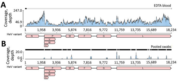
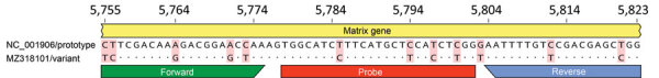
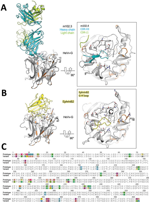
We identified and isolated a novel Hendra virus (HeV) variant not detected by routine testing from a horse in Queensland, Australia, that died from acute illness with signs consistent with HeV infection. Using whole-genome sequencing and phylogenetic analysis, we determined the variant had ≈83% nt identity with prototypic HeV. In silico and in vitro comparisons of the receptor-binding protein with prototypic HeV support that the human monoclonal antibody m102.4 used for postexposure prophylaxis and current equine vaccine will be effective against this variant. An updated quantitative PCR developed for routine surveillance resulted in subsequent case detection. Genetic sequence consistency with virus detected in grey-headed flying foxes suggests the variant circulates at least among this species. Studies are needed to determine infection kinetics, pathogenicity, reservoir-species associations, viral-host coevolution, and spillover dynamics for this virus. Surveillance and biosecurity practices should be updated to acknowledge HeV spillover risk across all regions frequented by flying foxes.

Keywords: Hendra henipavirus; Hendra virus; One Health; PCR; RNA sequencing; central nervous system; horse diseases; meningitis/encephalitis; sentinel species; sentinel surveillance; syncytia; syndromic surveillance; transdisciplinary research; vasculitis; viral encephalitis; viruses; whole-genome sequencing; zoonoses.


Figures

Figure 1
Figure 1
Sequence coverage of novel HeV variant from horse in Australia. The RNA sequencing reads were mapped to the novel HeV variant genome to examine coverage across the genome and depth for EDTA blood (A) and pooled swab samples (B). The x-axis shows the genome position with genes annotated and the y-axis shows the sequence read coverage (depth). Mean coverage depths were 46.9 for EDTA blood and 0.6 for pooled swab samples. V,W, and C indicate variably transcribed nonstructural proteins. F, fusion; G, glycoprotein; HeV, Hendra virus; M, matrix protein; N, nucleoprotein; P, phosphoprotein.
Figure 2
Figure 2
Genomic variation in the Hendra virus (HeV) matrix gene assay primer/probe binding sites for novel HeV variant from horse in Australia. The genomic region targeted by the commonly used HeV matrix gene quantitative RT-PCR assay (28) was aligned and compared for the prototype and variant HeV strains. The genomic positions relative to the prototype strain (GenBank accession no. NC_001906) are shown at the top. Primers (forward and reverse) and probe binding sites are indicated by the colored bars. Mismatches between the sequences are highlighted with red shading; dots indicate identical bases.
Figure 3
Figure 3
Transmission electron micrographs of Vero cells inoculated with the EDTA blood sample in study of novel Hendra virus variant from horse in Australia. A) Thin section showing inclusion body (IB) within the cytoplasm (C) of multinucleated (N1 and N2) syncytial cell. The nonmembrane bound IB consists of hollow nucleocapsids. B) Thin section showing virion (red arrow) with egress occurring at the plasma membrane. C) Negative contrast analysis shows a double-fringed envelope of the virion (red arrow). D) Negative contrast analysis shows strands of ribonucleic protein characteristic of the family Paramyxoviridae. Scale bars represent 100 nm.
Figure 4
Figure 4
Phylogenomics of novel HeV variant from horse in Australia. A) Maximum-likelihood phylogeny of paramyxoviruses using complete L protein sequences. Gray shading indicates henipaviruses, and red text indicates the novel HeV variant, which groups with the prototypic HeV. Bootstrap support values as proportions of 500 replicates are shown at nodes; values <0.7 are hidden. Scale bar indicates substitutions per site. B) Enlarged gray area from panel A shows branch lengths for the henipavirus clade. The branch leading back to the common ancestor of all known HeVs and the novel HeV variant does not exceed 0.03; thus, they are considered variants of the same species. C) Maximum-likelihood phylogeny of partial N and P where deep branch lengths have been collapsed for visualization only to demonstrate that the variant is well outside the known diversity of HeV. Scale bar indicates substitutions per site. D) Nucleotide genomic similarity of the variant compared with the prototypic HeV strain. V,W, and C indicate variably transcribed nonstructural proteins. F, fusion; G, glycoprotein; HeV, Hendra virus; L, paramyxovirus polymerase; M, matrix protein; N, nucleoprotein; P, phosphoprotein.
Figure 5
Figure 5
In silico modeling of HeV) from a horse in Australia. A, B) The translated protein sequence encoded by the HeV-var variant G gene was modeled using the known protein structure of the prototype virus bound to the human mAb m102.4 (A) and the receptor ephrinB2 (B). Side views (at left) of the interactions between the HeV G protein and the 2 binding partners highlight key binding residues (as sticks) and the variant positions (orange) relative to the m102.4 (heavy chain in teal and light chain in green) and ephrinB2 (in yellow). Zoomed top views (at right) of the HeV G and m102.4/ephrinB2 binding interface highlight specific interactions by the complementarity-determining regions of the mAb and G-H loop of the receptor ephrinB2. These data show that variable positions do not occur at critical epitopes at the HeV G and m102.4 binding interface and have very minor effect on the receptor ephrinB2 binding. C) Alignment of the prototypic and variant HeV strain G proteins. Variable positions are highlighted in color. F, fusion; G, glycoprotein; HeV, Hendra virus; mAb, monoclonal antibody.

References

    1. Eaton BT, Broder CC, Middleton D, Wang L-F. Hendra and Nipah viruses: different and dangerous. Nat Rev Microbiol. 2006;4:23–35. 10.1038/nrmicro1323 - DOI - PMC - PubMed
    1. Selvey LA, Wells RM, McCormack JG, Ansford AJ, Murray K, Rogers RJ, et al. Infection of humans and horses by a newly described morbillivirus. Med J Aust. 1995;162:642–5. 10.5694/j.1326-5377.1995.tb126050.x - DOI - PubMed
    1. Wong KT, Tan CT. Clinical and pathological manifestations of human henipavirus infection. Curr Top Microbiol Immunol. 2012;359:95–104. 10.1007/82_2012_205 - DOI - PubMed
    1. Playford EG, McCall B, Smith G, Slinko V, Allen G, Smith I, et al. Human Hendra virus encephalitis associated with equine outbreak, Australia, 2008. Emerg Infect Dis. 2010;16:219–23. 10.3201/eid1602.090552 - DOI - PMC - PubMed
    1. Murray K, Rogers R, Selvey L, Selleck P, Hyatt A, Gould A, et al. A novel morbillivirus pneumonia of horses and its transmission to humans. Emerg Infect Dis. 1995;1:31–3. 10.3201/eid0101.950107 - DOI - PMC - PubMed

Publication types