. 2022 Mar 3;82(5):950-968.e14.
doi: 10.1016/j.molcel.2022.02.011. Epub 2022 Feb 23.

Regulation of GTPase function by autophosphorylation

Affiliations

Regulation of GTPase function by autophosphorylation

Christian W Johnson et al. Mol Cell. .

Abstract

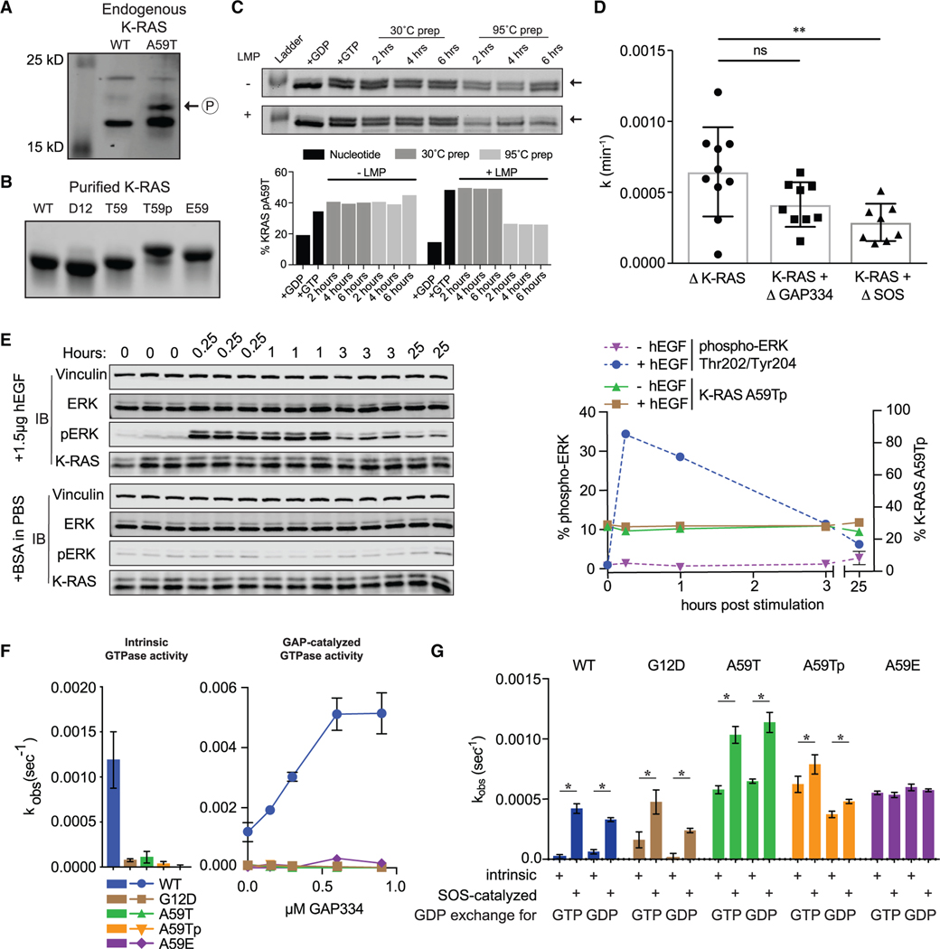
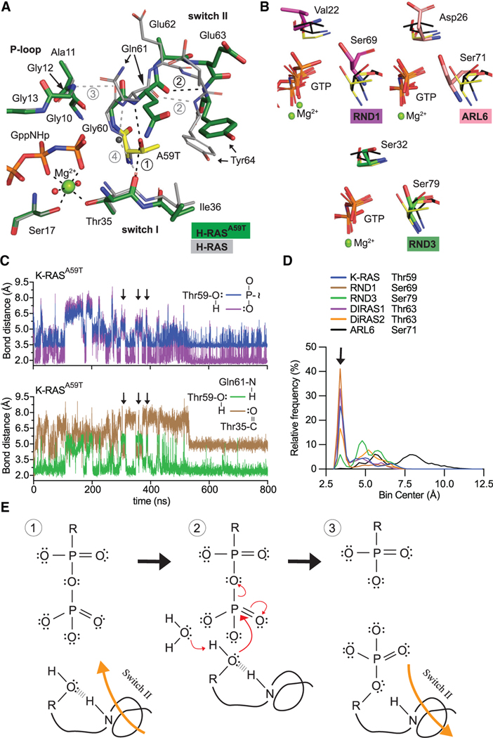
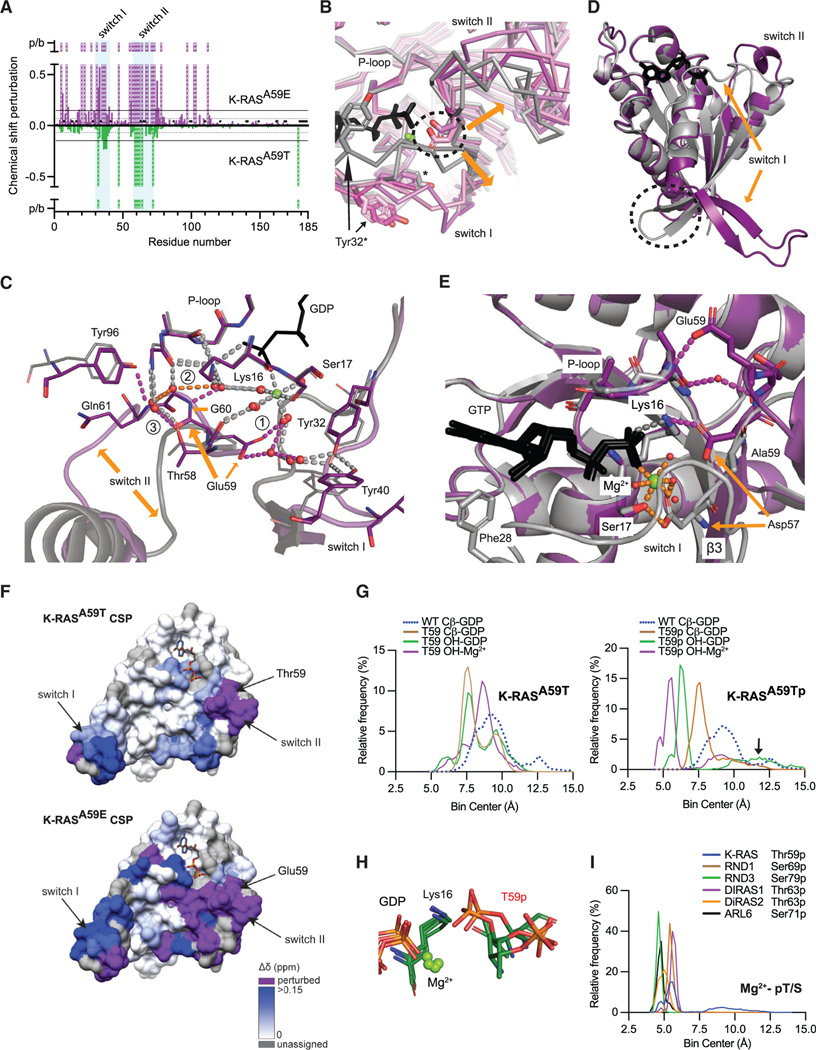
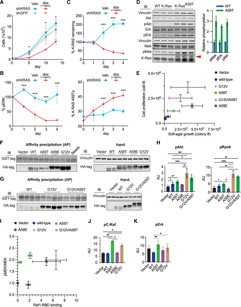
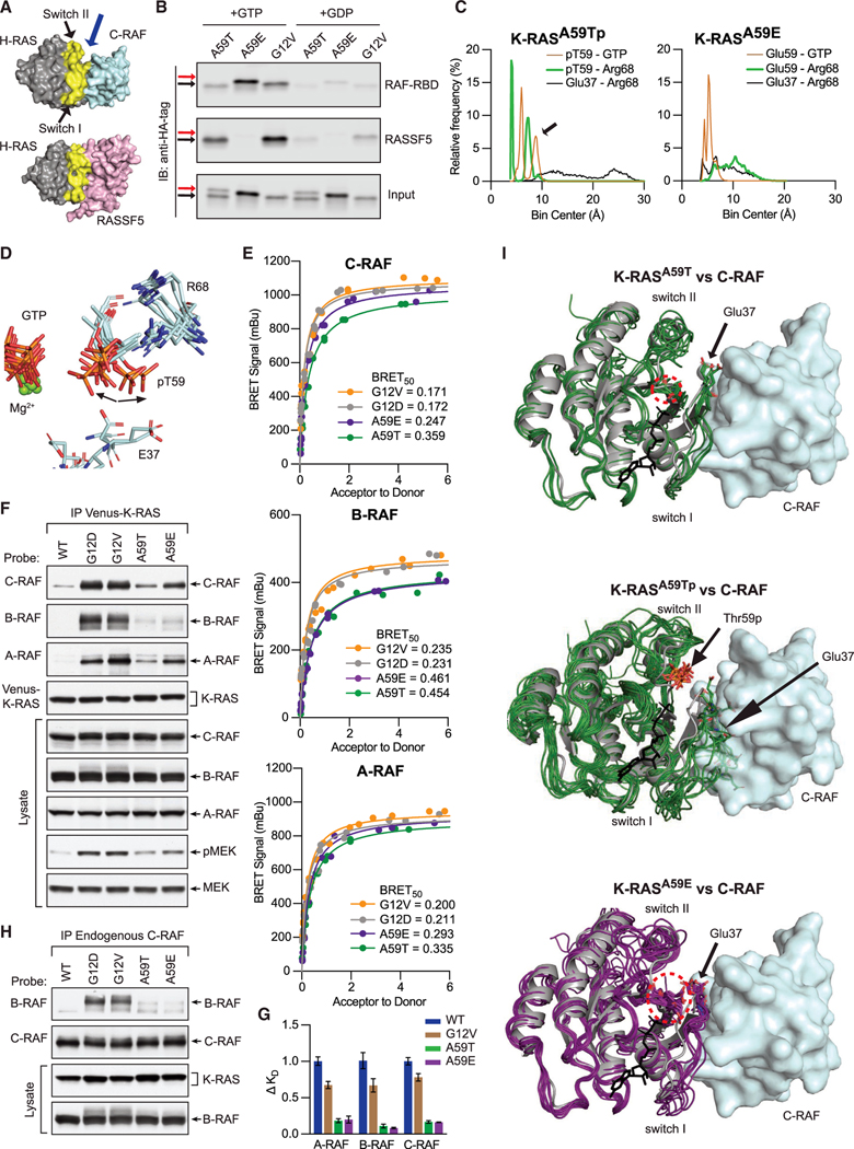
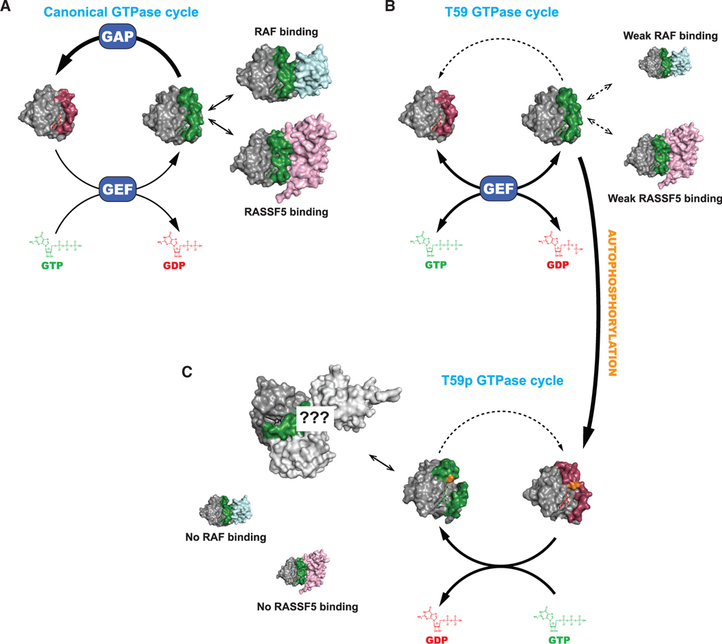
A unifying feature of the RAS superfamily is a conserved GTPase cycle by which these proteins transition between active and inactive states. We demonstrate that autophosphorylation of some GTPases is an intrinsic regulatory mechanism that reduces nucleotide hydrolysis and enhances nucleotide exchange, altering the on/off switch that forms the basis for their signaling functions. Using X-ray crystallography, nuclear magnetic resonance spectroscopy, binding assays, and molecular dynamics on autophosphorylated mutants of H-RAS and K-RAS, we show that phosphoryl transfer from GTP requires dynamic movement of the switch II region and that autophosphorylation promotes nucleotide exchange by opening the active site and extracting the stabilizing Mg2+. Finally, we demonstrate that autophosphorylated K-RAS exhibits altered effector interactions, including a reduced affinity for RAF proteins in mammalian cells. Thus, autophosphorylation leads to altered active site dynamics and effector interaction properties, creating a pool of GTPases that are functionally distinct from their non-phosphorylated counterparts.

Keywords: GTPase; NMR; RAF; RAS; RASSF; autophosphorylation; kinase; molecular dynamics; phosphoryl transfer; protein crystallography.


Conflict of interest statement

Declaration of interests K.M.H. is married to a member of Molecular Cell’s Advisory Board.

Figures

Figure 1.
Figure 1.. T59 phosphorylation alters K-RAS cycling
(A) Western blot of K-RAS in LIM1215 (WT) and SNU-175 (A59T) cells. (B) SDS-PAGE of purified K-RAS with different residues at position 59. (C) K-RASA59T incubated in the presence of GDP (lane 2) or GTP (lane 3). Dephosphorylation of K-RASA59T incubated with GTP with or without lambda phosphatase (LMP) for different times at 30°C after pre-incubation of protein at 30°C (lanes 4–6) or 95°C (lanes 7–9). Band quantification is shown below. (D) Summary of K-RASA59T autophosphorylation kinetics alone or with regulatory proteins. Autophosphorylation rate constants (k) were calculated as v•[E0]−1. (E) Serum-starved and hEGF-stimulated SNU-175 cells showing phosphorylated ERK1/2 (pERK) or K-RAS. Replicates are labeled above the gel. Band quantification was normalized to the average “0” replicate and is shown on the right. (F) Intrinsic and GAP-catalyzed hydrolysis (k) for K-RAS and mutants. Each bar or data point represents the average k (n = 3–4). (G) Exchange of GDP-loaded K-RAS4B for mant-GTP or mant-GDP (n = 3–4). * denotes p < 0.05 and ** denotes p < 0.005 by Student’s t test. Error bars represent ± SD.
Figure 2.
Figure 2.. Conserved mechanism of autophosphorylation in GTPases
(A) Active site comparison of H-RASA59T crystal 1 (green) and WT H-RAS (PDB: 3K8Y, gray). Black and gray dashed lines are H-bonds made in H-RASA59T and WT structures. Thr59 is shown in yellow. (B) Active site similarities between H-RASA59T and other small GTPases. Black sticks are from the H-RASG12V/A59T structure (PDB: 521P) with an alternate Thr59 orientation. (C) Bond distances made during simulation of K-RASA59T bound to GTP. Inset on the right shows measured bonds. (D) Frequency distribution of nucleophile to γ-phosphate distances from MD simulations. (E) Proposed mechanism of autophosphorylation. The N-H group of switch II represents the backbone carbonyl of Gln61.
Figure 3.
Figure 3.. The molecular mechanism of hyperexchange
(A) Chemical shift perturbations induced by mutation of Ala59 plotted by K-RAS residue. 1H-15N cross-peaks for K-RASA59E or K-RASA59T are referenced to WT K-RAS-GppNHp. K-RASA59T versus WT chemical shift perturbations are shown as negative values to compare changes to K-RASA59E. Resonances detected in WT, but not in mutants, due to perturbations or severe peak broadening (p/b), are shown as dashed bars with an arbitrary value of 1 ppm. Residues not assigned for WT are marked with X. Horizontal lines show threshold of mean chemical perturbation plus one SD for K-RASA59T (gray, dashed) and K-RASA59E (solid black). (B) Comparison of K-RASA59E crystal structures bound to GppNHp (purple) and GDP (pink) to WT H-RAS bound to GDP or GppNHp (gray). Dashed circle indicates Glu59. (C) Molecule B of H-RASA59E bound to GDP (purple). Glu59 rearranges the active site to favor GDP release, unlike the WT reference structure (PDB: 4OBE, gray). Gray dashes are shared H-bonds between structures. Colored dashes are described in text. (D) Structure of GDP-bound K-RASA59E lacking Mg2+ compared to the WT reference structure. Dashed circle shows junction between switch I and helix 1. (E) Glu59 and Asp57 stabilize a pro-exchange active site in K-RASA59E. (F) Chemical shift perturbations (A) were mapped to the K-RAS surface. Color intensity represents magnitude of chemical shift changes relative to WT as defined by the scale. Resonances detected in WT, but not in the mutants, due to perturbation or broadening are shown in purple. Residues without assignments are colored in gray. (G) Frequency distribution of bond distances during MD simulations of K-RASA59T and K-RAST59p bound to GDP. (H) Cluster analysis of K-RAST59p MD simulation. (I) Frequency distribution of bond distances during MD simulations of phosphorylated GTPases bound to GDP.
Figure 4.
Figure 4.. Roles of K-RASA59T and K-RASA59E in cellular transformation
(A) Effect of KRAS shRNA on proliferation in SNU-175 cells (n = 2–3). (B) Effect of KRAS shRNA on ERK1/2 phosphorylation in SNU-175 cells (n = 2–3). Data are normalized to the average ERK1/2 phosphorylation on day 0. (C) Effect of KRAS shRNA on K-RAS expression and autophosphorylation in SNU-175 cells (n = 2–3). Data are normalized to the average K-RAS expression on day 0 (upper). (D) Baseline phosphorylation of Mek, Erk, and Akt in WT and K-RasA59T mouse embryonic stem cells after 6 h of serum starvation. The red arrow denotes autophosphorylated K-RAS. Quantification is shown on the right and normalized to average phosphorylation for WT cells. (E) Growth of NIH3t3 cells with ectopic K-RAS on plastic (x axis, n = 4–5) and in soft agar (y axis, n = 3) in 10% serum. (F and G) Representative western blots from precipitation of GTP-bound K-RAS from NIH3t3 cells (F and G) by C-RAF-RBD-GST. Note that the upper band representing K-RAST59p is absent. (H) Quantification of Akt (Ser473) and Rps6 (Ser235/236) phosphorylation (n = 4). (I) Comparison of Mek phosphorylation (x axis, n = 4) and K-RAS-GTP (y axis, n = 4). Phosphorylated was normalized to total Mek. K-RAS-GTP was scaled to protein input and normalized to the average in cells expressing WT K-RAS. (J) Quantification of C-Raf phosphorylation on Ser289/296/301 (n = 4). (K) Quantification of Erk1/2 phosphorylation on Thr202/Tyr204 (n = 4). In (A–C), errors bars represent ± SD and statistical analyses used Student’s t test. For (H–K), errors bars represent ± SD and statistical analyses were performed with Mann-Whitney test. *, **, and *** represent P values of < 0.05, < 0.005, and < 0.0005, respectively. In (E–K), statistical comparisons and representative western blots are in Figure S4.
Figure 5.
Figure 5.. GTP induces phosphoryl movement to discriminate effector interactions
(A) Binding interfaces of C-RAF-RBD (PDB: 4G0N) and RASSF5-RBD (PDB: 3DDC). Blue arrows indicate Ala59 of RAS. (B) Pull-down of ectopic K-RAS, preloaded with GTP or GDP, by C-RAF-RBD-GST or RASSF5-GST. Arrows denote non-phosphorylated (black) and phosphorylated (or A59E) (red) K-RAS. (C) Frequency distribution of bond distances during MD simulations of mutant K-RAS bound to GTP. (D) MD simulation cluster analysis of K-RAST59p. (E) BRET saturation curves showing interaction between RAF isoform regulatory domains and K-RAS mutants in HEK293FT cells. (F) Western blot of RAF isoforms co-immunoprecipitating with Venus-tagged K-RAS from serum-starved HeLa cells. (G) Affinity changes of K-RAS for the RAF-RBDs determined by BLI. Data normalized to WT K-RAS (KDWT/KDMut). Error bars represent ± SD (n = 2–4) (H) Co-immunoprecipitation of B-RAF by C-RAF with different K-RAS mutants from serum-starved HeLa cells. (I) Comparison of K-RAS conformations (green, purple) generated by cluster analysis of MD simulations to H-RAS (gray) bound to C-RAF (PDB: 4G0N, cyan). Nucleotide is shown in black.
Figure 6.
Figure 6.. Influence of autophosphorylation on nucleotide cycling and effector engagement
(A) The normal GTPase cycle of K-RAS. (B) Thr59 inhibits nucleotide hydrolysis and promotes intrinsic nucleotide exchange, activating K-RAS. Subsequently, the mutant shows weak binding to RAF and RASSF5 proteins. (C) When Thr59 becomes phosphorylated (orange), K-RAS enters an alternative cycle where it becomes insensitive to GEF and loses the ability to bind to RAF and RASSF5 but opens up the possibility of interacting with novel effectors.

References

    1. Abraham MJ, Murtola T, Schulz R, Páll S, Smith JC, Hess B, and Lindahl E. (2015). GROMACS: High performance molecular simulations through multi-level parallelism from laptops to supercomputers. SoftwareX 1-2, 19–25.
    1. Adams PD, Afonine PV, Bunkóczi G, Chen VB, Davis IW, Echols N, Headd JJ, Hung LW, Kapral GJ, Grosse-Kunstleve RW, et al. (2010). PHENIX: a comprehensive Python-based system for macromolecular structure solution. Acta Crystallogr. D Biol. Crystallogr 66, 213–221. - PMC - PubMed
    1. Ahearn I, Zhou M, and Philips MR (2018). Posttranslational Modifications of RAS Proteins. Cold Spring Harb. Perspect. Med 8, a031484. - PMC - PubMed
    1. Barthelmes K, Ramcke E, Kang HS, Sattler M, and Itzen A. (2020). Conformational control of small GTPases by AMPylation. Proc. Natl. Acad. Sci. USA 117, 5772–5781. - PMC - PubMed
    1. Bergom C, Hauser AD, Rymaszewski A, Gonyo P, Prokop JW, Jennings BC, Lawton AJ, Frei A, Lorimer EL, Aguilera-Barrantes I, et al. (2016). The tumor-suppressive small GTPase DiRas1 binds the noncanonical guanine nucleotide exchange factor SmgGDS and antagonizes SmgGDS interactions with oncogenic small GTPases. J. Biol. Chem 291, 10948. - PMC - PubMed

Publication types