. 2022 Feb 9;11(4):597.
doi: 10.3390/cells11040597.

Anti-Aging Effects of a Serum Based on Coconut Oil Combined with Deer Antler Stem Cell Extract on a Mouse Model of Skin Aging

Affiliations

Anti-Aging Effects of a Serum Based on Coconut Oil Combined with Deer Antler Stem Cell Extract on a Mouse Model of Skin Aging

Truc Le-Buu Pham et al. Cells. .

Abstract

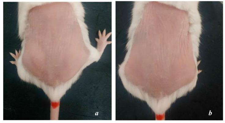
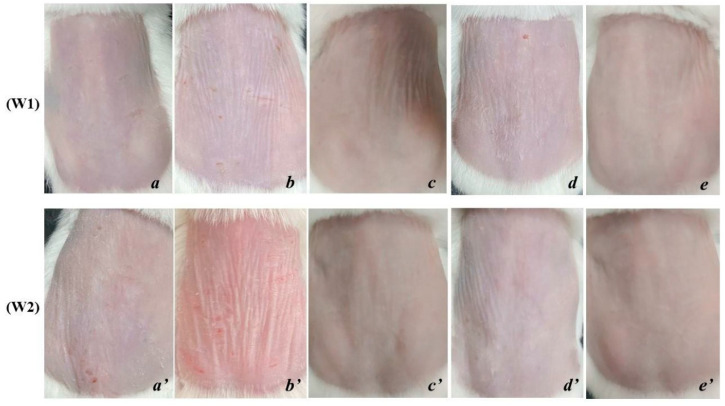
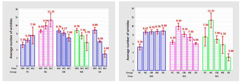
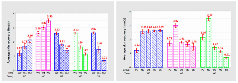
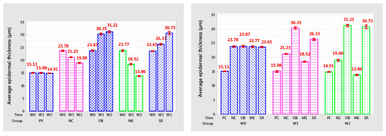
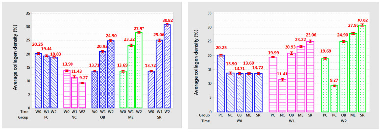
Anti-aging is one of the top goals in the field of health care and aesthetics. Anti-aging cosmetics derived from nature are oriented to long-term development, bringing safety to users and being environmentally friendly. The aim of this study was to develop an anti-aging cosmetic formulation process based on coconut oil in combination with deer antler stem cell extract. The results show that the presence of deer antler stem cell extract added to the foundation made the serum product highly stable and helped improve skin aging significantly after 2 weeks of use. The skin site where the serum product was applied showed a smooth and elastic skin surface, with very few fine lines and shallow wrinkles. Serum reduced the number of wrinkles (48.09% compared to commercial serum (ME) and 60.31% compared to positive control (PC)), reduced skin recovery time (39.31% compared to ME and 67.1% of PC) after two weeks of use. After 2 weeks of use, collagen density increased 10.18% compared to ME and 63.76% compared to control. Epidermal thickness increased by 106.1% compared to PC and 121.7% compared to ME.

Keywords: deer antler stem cells; mouse; ultraviolet A (UVA); virgin coconut oil (VCO).


Conflict of interest statement

The authors declare no competing interest.

Figures

Figure 1
Figure 1
Mouse skin after 8 weeks of UVA exposure: ((a)—PC; (b)—NC).(a)The PC group showed many statistically significant superficial wrinkles.(b) NC group dry skin, uneven skin tone, deep wrinkles and a lot.
Figure 2
Figure 2
Mouse skin after 1 and 2 weeks of applying the experimental product (W1—after 1 week; W2—after 2 weeks; (a,a’)—PC; (b,b’)—NC; (c,c’)—OB; (d,d’)—ME; (e,e’)—SR). (a) PC, wrinkles are more and more obvious, (a’) wrinkles are more and deeper. (b) NC, many and deep wrinkles; (b’); dry, vulnerable skin, many and deep wrinkles. (c) OB, smooth, even skin tone, few wrinkles but deep; (c’)—smooth skin, few and shallow wrinkles. (d) ME, dry skin, many and shallow wrinkles; (d’)—few wrinkles, shallow, smooth skin. (e) SR, skin color is relatively uniform, smooth and with few wrinkles; (e’)—smooth skin, few or no wrinkles.
Figure 3
Figure 3
The graph shows the average number of wrinkles after each week of product application.
Figure 4
Figure 4
The graph shows the average number of skin recovery times after each week of applying the product.
Figure 5
Figure 5
Mouse skin microstructure after 8 weeks of UVA (X20) irradiation ((a)—PC; (b)—NC). E: epidermis; D: dermis; PD: papillary dermis ; CB: collagen bundles; SG: sebaceus glance; SC: stratum corneum; BV: blood vessels ; HF: hair folicle; FB: fibroplast [37].
Figure 6
Figure 6
Trichrome-stained mouse skin structure after 2 weeks of treatment (X20): (W1—after 1 week; W2—after 2 weeks; (a,a’)—PC; (b,b’)—NC; (c,c’)—OB; (d,d’)—ME; (e,e’)—SR). (a,a’)—PC, very little difference, relatively similar to the time before applying the product. (b,b’)—NC, discrete fiber bundles, almost no collagen association, low collagen density. (c,c’)—OB, collagen structure is more interconnected, cell proliferation and a small amount of new blood vessels, epidermis still increases in thickness and tends to be relatively keratinized. (d,d’)—ME, relatively high collagen density, appearance of new cells and blood vessels under the skin. (e,e’)—SR, dense, connective collagen density, high number of secretory cells, hair follicle cells and new blood vessels.
Figure 7
Figure 7
The graph shows the average epidermal thickness of the mouse skin after each week of treatment.
Figure 8
Figure 8
The graph shows the average collagen density (%) of the mouse skin after each week of treatment.

References

    1. Sjerobabski-Masnec I., Šitum M. Starenje kože lica. Acta Clin. Croat. 2010;49:515–518. - PubMed
    1. Baumann L. Skin ageing and its treatment. J. Pathol. 2007;211:241–251. doi: 10.1002/path.2098. - DOI - PubMed
    1. Krutmann J., Bouloc A., Sore G., Bernard B.A., Passeron T. The skin aging exposome. J. Dermatol. Sci. 2017;85:152–161. doi: 10.1016/j.jdermsci.2016.09.015. - DOI - PubMed
    1. Puizina-Ivic N. Skin aging. Acta Dermatovenerol. Alp. Panon. Adriat. 2008;17:47. - PubMed
    1. Uitto J. Understanding Premature Skin Aging. N. Engl. J. Med. 1997;337:1463–1465. doi: 10.1056/NEJM199711133372011. - DOI - PubMed

Publication types