. 2022 Feb 10;11(4):609.
doi: 10.3390/cells11040609.

Calcineurin Controls Cellular Prion Protein Expression in Mouse Astrocytes

Affiliations

Calcineurin Controls Cellular Prion Protein Expression in Mouse Astrocytes

Giulia Dematteis et al. Cells. .

Abstract

Prion diseases arise from the conformational conversion of the cellular prion protein (PrPC) into a self-replicating prion isoform (PrPSc). Although this process has been studied mostly in neurons, a growing body of evidence suggests that astrocytes express PrPC and are able to replicate and accumulate PrPSc. Currently, prion diseases remain incurable, while downregulation of PrPC represents the most promising therapy due to the reduction of the substrate for prion conversion. Here we show that the astrocyte-specific genetic ablation or pharmacological inhibition of the calcium-activated phosphatase calcineurin (CaN) reduces PrPC expression in astrocytes. Immunocytochemical analysis of cultured CaN-KO astrocytes and isolation of synaptosomal compartments from the hippocampi of astrocyte-specific CaN-KO (ACN-KO) mice suggest that PrPC is downregulated both in vitro and in vivo. The downregulation occurs without affecting the glycosylation of PrPC and without alteration of its proteasomal or lysosomal degradation. Direct assessment of the protein synthesis rate and shotgun mass spectrometry proteomics analysis suggest that the reduction of PrPC is related to the impairment of global protein synthesis in CaN-KO astrocytes. When WT-PrP and PrP-D177N, a mouse homologue of a human mutation associated with the inherited prion disease fatal familial insomnia, were expressed in astrocytes, CaN-KO astrocytes showed an aberrant localization of both WT-PrP and PrP-D177N variants with predominant localization to the Golgi apparatus, suggesting that ablation of CaN affects both WT and mutant PrP proteins. These results provide new mechanistic details in relation to the regulation of PrP expression in astrocytes, suggesting the therapeutic potential of astroglial cells.

Keywords: FK506; astrocytes; calcineurin; calcium; neuroinflammation; prion protein; protein synthesis.


Conflict of interest statement

The authors declare no conflict of interest.

Figures

Figure 1
Figure 1
Analysis of PrPC expression in Ctr, FK506-treated Ctr and CaN-KO primary hippocampal astrocytes. (a) Expression levels of PrPC in Ctr and CaN-KO astrocytes. WB analysis of protein extracts with anti-PrPC 12B2 and anti-actin antibodies and quantification of actin-normalized PrPC levels expressed as percentages of Ctr. Data are the means ± SEM of Ctr (n = 4) and KO (n = 6) replicate astrocyte cultures. *** p < 0.001 by unpaired t-test. (b) Deglycosylation assay in Ctr and CaN-KO astroglial lysates. Ctr and CaN-KO astroglial lysates were treated with PNGase F and analysed by WB with anti-PrP 12B2 and anti-actin antibodies. (c) Time-course analysis with FK506 in Ctr and CaN-KO astrocytes. Ctr astrocytes were left untreated or treated with FK506 200 nM for 7 days and analyzed by WB with anti-PrP 12B2 and anti-actin antibodies, along with CaN-KO astrocyte lysates. (d) Quantification of actin-normalized PrPC levels in WB like the one shown in (c). Data are the means ± SEM, (n = 6–11); * p < 0.05, ** p < 0.01 vs. Ctr by one-way Anova, multiple comparison Kruskal–Wallis test.
Figure 2
Figure 2
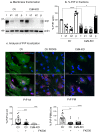
Analysis of PrPC localization in Ctr, FK506-treated Ctr and CaN-KO primary hippocampal astrocytes. (a) Fractionation of Ctr and CaN-KO astrocytic lysates by centrifugation and evaluation of PrPC relative abundance by WB with anti-PrP in t (total cell lysates), s1 (post nuclear supernatant), s2 (soluble fraction), p (membrane fraction) fractions. (b) Quantification of membrane fractionation is expressed as means ± SEM, n = 5; **** p < 0.0001, * p < 0.05 by one-way Anova, Sidak’s multiple comparison. (c) Immunofluoresce analysis on Ctr, FK506-treated Ctr and CaN-KO astrocytes with anti-PrP (green), anti-BAP31 (pink) antibodies and reacted with DAPI to stain the nuclei (blue). Confocal microscope analysis, scale bar 20 µm. Quantification of total PrP (PrP tot) or plasma membrane PrP (PrP PM) fluorescence density expressed as percentages of Ctr from Ctr n = 54 cells, FK506-treated Ctr n = 67 and CaN-KO n = 70 cells, from four to five replicates, ** p < 0.01, *** p < 0.001 and **** p < 0.0001.
Figure 3
Figure 3
Analysis of PrPC expression in hippocampal tissues from ACN-Ctr and ACN-KO mice at one month of age. (a) WB analysis with anti-PrP and anti-actin antibodies of hippocampal homogenates from ACN-Ctr and ACN-KO mice. Quantification of the actin-normalized PrP signal expressed as percentage of Ctr. Data are the means ± SEM, ACN-Ctr n = 7, ACN-KO n = 8, unpaired t-test, ns. (b) Synaptosomal fractions from hippocampi of ACN-Ctr and ACN-KO mice were analyzed by WB with anti-PrP and anti-actin antibodies. Quantification of the actin-normalized PrP signal expressed as percentage of Ctr. Data are the means ± SEM, ACN-Ctr n = 4, ACN-KO n = 4, unpaired t-test, ** p = 0.0015. (c) WB analysis with anti-PrP and anti-actin antibodies of hippocampal neurons from ACN-Ctr (Ctr) and ACN-KO (CaN-KO) mice. Quantification of the actin-normalized PrP signal expressed as percentages of Ctr. Data are the means ± SEM, Ctr n = 6, CaN-KO n = 8, unpaired t-test, ns.
Figure 4
Figure 4
Real-time PCR, proteasomal degradation analysis of Ctr, FK506-treated Ctr and CaN-KO astrocytes. (a) Real-time PCR of Prnp on primary astrocytes, Ctr, FK506 (200 nM for 7 days)-treated and CaN-KO. Values represent means ± SEM ∆C(t) of gene/S18 of five independent experiments for each condition. (b) WB analysis of PrPC and actin, protein degradation in hippocampal astrocytes from Ctr, FK506 (200 nM for 7 days)-treated and CaN-KO. Where indicated, MG132 was added 3 h before lysis. Data are expressed as means ± SEM, three independent cultures were used, one-way Anova, multiple comparison, * p < 0.05, *** p < 0.001 and **** p < 0.0001.
Figure 5
Figure 5
Analysis of lysosomal-dependent PrPC degradation. Ctr and CaN-KO astrocytes were untreated or treated with chloroquine (CQ) before immunofluorescence with anti PrP (green), BAP31 (pink) antibodies and reacted with DAPI to stain the nuclei (blue). Confocal microscope analysis, scale bar 20 µm. Quantification of total PrPC fluorescence density expressed as percentage of Ctr. Data are the means ± SEM of Ctr n = 49 cells, Ctr CQ n = 46 and KO n = 58 cells; KO CQ = 62, from three to seven coverslips from three independent experiments, * p < 0.05.
Figure 6
Figure 6
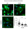
Protein synthesis analysis in Ctr, FK506-treated Ctr and CaN-KO astrocytes. Ctr, FK506-treated Ctr and CaN-KO astrocytes were pulsed with 4 µM puromycin, fixed and analysed by IF with anti-puromycin antibody (green) and reacted with DAPI to stain the nuclei (blue). Images were acquired with a Leica Thunder imager 3D live cell microscope, scale bar 41.6 µm. Data are expressed as means ± SEM of n cells Ctr = 37, FK506-treated Ctr = 44, CaN-KO = 24, from three independent experiments. one-way Anova, Dunnett’s multiple comparison analysis, *** p < 0.001 and **** p < 0.0001.
Figure 7
Figure 7
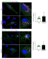
Analysis of WT and D177N PrP transient expression in Ctr and CaN-KO astrocytes. Ctr and CaN-KO astrocytes were transfected with eGFP-WT PrP (a) and eGFP-D177N PrP (b) fusion proteins (green), immunostained with an anti-GM130 (pink) to mark the Golgi apparatus and reacted with DAPI to stain the nuclei (blue). Images were acquired with a confocal microscope, scale bar 20 µm. PrP fluorescent density in the Golgi apparatus is expressed as mean ± SEM, from four independent coverslips, * p < 0.05 and ** p < 0.01.

References

    1. Prusiner S.B. Prions. Proc. Natl. Acad. Sci. USA. 1998;95:13363–13383. doi: 10.1073/pnas.95.23.13363. - DOI - PMC - PubMed
    1. Puoti G., Bizzi A., Forloni G., Safar J.G., Tagliavini F., Gambetti P. Sporadic Human Prion Diseases: Molecular Insights and Diagnosis. Lancet Neurol. 2012;11:618–628. doi: 10.1016/S1474-4422(12)70063-7. - DOI - PubMed
    1. Sy M.-S., Gambetti P., Wong B.-S. Human Prion Diseases. Med. Clin. N. Am. 2002;86:551–571. doi: 10.1016/S0025-7125(02)00004-4. - DOI - PubMed
    1. Takada L.T., Kim M.-O., Metcalf S., Gala I.I., Geschwind M.D. Chapter 29—Prion Disease. In: Geschwind D.H., Paulson H.L., Klein C., editors. Handbook of Clinical Neurology. Volume 148. Elsevier; Amsterdam, The Netherlands: 2018. pp. 441–464. Neurogenetics, Part II. - PubMed
    1. Marín-Moreno A., Espinosa J.C., Torres J.M. Transgenic Mouse Models for the Study of Prion Diseases. Prog. Mol. Biol. Transl. Sci. 2020;175:147–177. doi: 10.1016/bs.pmbts.2020.08.007. - DOI - PubMed

Publication types