. 2022 Feb 10;11(4):610.
doi: 10.3390/cells11040610.

MG132 Induces Progerin Clearance and Improves Disease Phenotypes in HGPS-like Patients' Cells

Affiliations

MG132 Induces Progerin Clearance and Improves Disease Phenotypes in HGPS-like Patients' Cells

Karim Harhouri et al. Cells. .

Abstract

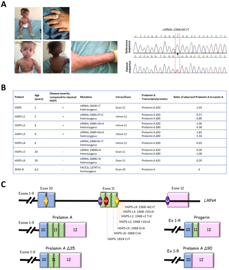
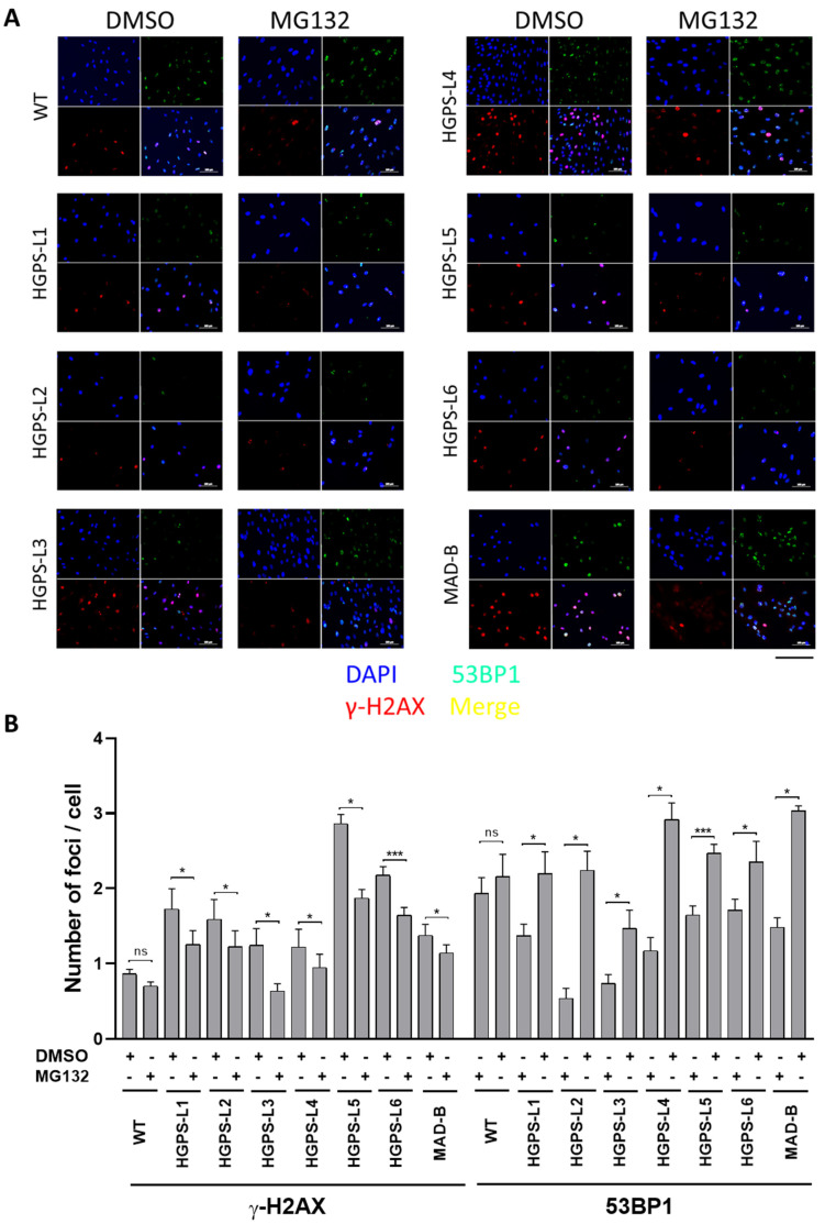
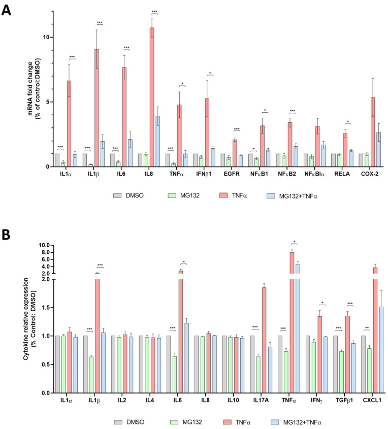
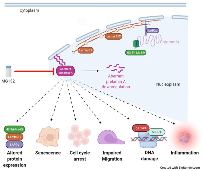
Progeroid syndromes (PS), including Hutchinson-Gilford Progeria Syndrome (HGPS), are premature and accelerated aging diseases, characterized by clinical features mimicking physiological aging. Most classical HGPS patients carry a de novo point mutation within exon 11 of the LMNA gene encoding A-type lamins. This mutation activates a cryptic splice site, leading to the production of a truncated prelamin A, called prelamin A ∆50 or progerin, that accumulates in HGPS cell nuclei and is a hallmark of the disease. Some patients with PS carry other LMNA mutations and are named "HGPS-like" patients. They produce progerin and/or other truncated prelamin A isoforms (∆35 and ∆90). We previously found that MG132, a proteasome inhibitor, induced progerin clearance in classical HGPS through autophagy activation and splicing regulation. Here, we show that MG132 induces aberrant prelamin A clearance and improves cellular phenotypes in HGPS-like patients' cells other than those previously described in classical HGPS. These results provide preclinical proof of principle for the use of a promising class of molecules toward a potential therapy for children with HGPS-like or classical HGPS.

Keywords: MAD-B; MG132; autophagy; inflammation; prelamin A Δ35; prelamin A Δ90; progeria-like; progerin.


Conflict of interest statement

The authors declare no competing or conflicting interests.

Figures

Figure 1
Figure 1
Clinical and molecular description of cell lines. (A) Pictures and electropherograms of patient HGPS-L4 at age 6 years showing progeroid features, including sparse hair and eyebrows, small chin, a thin nose, prominent nipples, dyspigmentation with hyper/hypo-pigmented areas, and sclerodermatous changes on her chest. The heterozygous c.1968+6C>T LMNA mutation was confirmed by Sanger sequencing. (B) Characterization of LMNA and FACE1 (ZMPSTE24) gene mutations in HGPS-like and MAD-B patients eliciting aberrant prelamin A splicing or wild-type prelamin A accumulation. Variable disease severities compared to classical HGPS are indicated with “+”: more, “− “: less, or “=”: equal severity. The ratios of aberrant prelamin A to lamin A isoforms are shown, issued from Western blot data, except for prelamin A, against which no antibodies are available, and so were determined based on the transcript levels. (C) Locations of LMNA mutations and schematic representation of the aberrant prelamin A isoforms. SD: splice donor site, SA: splice acceptor site.
Figure 2
Figure 2
MG132 reduces aberrant prelamin A levels in HGPS-like and MAD-B fibroblasts. (A) Downregulation of aberrant prelamin A transcripts (Δ50: progerin, Δ35, Δ90, and WT prelamin A) in response to MG132. Quantitative real-time PCR analyses of lamin A, prelamin A Δ50 (progerin), prelamin A Δ35, prelamin A Δ90, lamin C, and RPS13 mRNA levels in HGPS, HGPS-like, MAD-B, and WT fibroblasts treated for 24 h with 500 nM MG132 relative to DMSO-treated cells (Control). The fold change of each transcript was determined by normalizing its value to that of RPS13 for each condition. (mean ± SEM, n = 4, Student’s t-test, * p < 0.05, ** p < 0.01, experimental vs. control). (B) MG132 reduced aberrant prelamin A protein levels in HGPS-like and MAB-B patient cells. Upper panels: Western blotting evaluation of lamin A/C, progerin, prelamin A, prelamin A ∆35 in whole-cell lysates from WT, HGPS, HGPS-like, and MAD-B fibroblasts treated with DMSO (−), 500 nM MG132 for 48 h (+). Lower panels: lamin A/C, progerin, prelamin A, prelamin A ∆35 expression levels were normalized to GAPDH values using ImageJ software. (mean ± SEM, n = 3, Student’s t-test, * p < 0.05, ** p < 0.01, *** p < 0.001. MG132-treated vs. DMSO-treated cells).
Figure 3
Figure 3
MG132 reduces senescence and enhances proliferation and migration in HGPS-like and MAD-B patients’ cells. (A) Luminescence-based quantification of senescence rate in WT, HGPS-like, and MAD-B fibroblasts treated with 500 nM MG132 for 96 h relative to DMSO-treated cells. Each experiment was performed on cells at the same passage level. Senescence is determined as relative light units (RLUs). (mean ± SEM, n = 3, Student’s t-test, * p < 0.05, ** p < 0.01. MG132-treated vs. DMSO-treated cells). (B) Colorimetric detection of senescence-associated β galactosidase in HGPS-like and MAD-B fibroblasts treated with 500 nM MG132 for 96 h relative to DMSO-treated cells. Each experiment was performed on cells at the same passage level. β-galactosidase blue staining is lower in cells treated with MG132 compared to cells treated with DMSO. (C) Cell proliferation rate based on the incorporation of bromodeoxyuridine (BrdU) into the DNA was expressed as absorbance OD 450 nm. (D) Left panel: an example of wound healing assay performed on HGPS-L1 fibroblasts treated for 6 h with DMSO or MG132 (500 nM). Right panel: the results of wound healing assays on individual samples (Figure S1) were grouped into biological replicates (1 HGPS, 6 HGPS-like, and 1 MAD-B) to perform statistical tests. (mean ± SEM, n = 8, Student’s t-test, *** p < 0.001. MG132-treated vs. DMSO-treated cells). Results are expressed as a percentage of the area of the original wound and normalized to DMSO-treated cells, considered as 100%. Scale bar, 100 µm.
Figure 4
Figure 4
MG132 treatment rescues the level of proteins, the expression of which is altered in HGPS-like and MAD-B cells. (A) Immunofluorescence microscopy on primary dermal fibroblasts from a healthy individual (WT) and HGPS-like and MAD-B patients, treated with 500 nM MG132 or an equal volume of DMSO for 48 h. Cells were stained with DAPI (blue) and antibodies to tri-methyl lysine 9 of core histone H3 (H3-Tri-Me-k9), lamin B1, and LAP2α. (B) Fold change in signal intensity in DMSO-treated patient’s cells relative to DMSO-treated WT cells, each normalized to the corresponding nuclei number. The signal intensity was set to 1 in DMSO-treated WT cells (mean ± SEM, n = 3, Student’s t-test, * p < 0.05, ** p < 0.01, *** p < 0.001). (C) Fold change in signal intensity in MG132-treated relative to DMSO-treated HGPS fibroblasts, each normalized to the corresponding nuclei number. The signal intensity was set to 1 in DMSO-treated cells. At least 200 fibroblast nuclei were randomly selected for each cell line (mean ± SEM, n = 3, Student’s t-test, * p < 0.05, ** p < 0.01, *** p < 0.001) and examined using ImageJ software. Scale bar, 200 µm.
Figure 5
Figure 5
Treatment of HGPS-like and MAD-B cells with MG132 reduces the levels of DNA damage. (A) Immunofluorescence microscopy on primary dermal fibroblasts from a healthy individual (WT) and HGPS-like and MAD-B patients, treated with 500 nM MG132 or an equal volume of DMSO for 48 h. Cells were stained with DAPI (blue) and antibodies to the indicated proteins. Scale bar, 200 µm. (B) Quantification of the number of foci of γ-H2AX and 53BP1 per cell in MG132-treated fibroblasts compared to DMSO-treated fibroblasts (mean ± SEM, n = 3, Student’s t-test, * p < 0.05, *** p < 0.001).
Figure 6
Figure 6
Anti-inflammatory effects of MG132 in HGPS-like and MAD-B cells. (A) Quantitative real-time PCR using selected inflammatory genes expression arrays in culture supernatants of HGPS, HGPS-like, and MAD-B fibroblasts treated for 6 h with MG132 (500 nM), TNFα (10 ng/mL) alone and in combination, or DMSO as vehicle control. The results of individual samples (Figure S5) were grouped into biological replicates (1 HGPS, 6 HGPS-like, and 1 MAD-B) to perform statistical tests. (mean ± SEM, n = 8, Student’s t-test, * p < 0.05, *** p < 0.001; MG132-treated vs. DMSO-treated cells and MG132+TNFα-treated vs. MG132-treated cells). (B) Enzyme-linked immunosorbent assay (ELISA) using multi-analyte ELISA arrays to measure inflammatory cytokines in culture supernatants from HGPS, HGPS-like, and MAD-B fibroblasts treated for 24 h with MG132 (500 nM), TNFα (10 ng/mL) alone and in combination, or DMSO as vehicle control. The results of individual samples (Figure S6) were grouped into biological replicates (1 HGPS, 6 HGPS-like, and 1 MAD-B) to perform statistical tests. (mean ± SEM, n = 8, Student’s t-test, * p < 0.05, ** p < 0.01, *** p < 0.001; MG132-treated vs. DMSO-treated cells and MG132+TNFα-treated vs. MG132-treated cells).
Figure 7
Figure 7
Schema summarizing the results of the current study. MG132 reduces the transcript levels of the aberrant prelamin A isoforms and the corresponding proteins already expressed. MG132 treatment improves several pathological parameters, including altered protein expression, cellular senescence, cell cycle arrest, impaired migration, DNA damage, as well as inflammatory cytokine expression.

References

    1. Hennekam R.C. Hutchinson-Gilford progeria syndrome: Review of the phenotype. Am. J. Med. Genet. A. 2006;140:2603–2624. doi: 10.1002/ajmg.a.31346. - DOI - PubMed
    1. Gordon L.B., Massaro J., D’Agostino R.B., Sr., Campbell S.E., Brazier J., Brown W.T., Kleinman M.E., Kieran M.W. Impact of farnesylation inhibitors on survival in Hutchinson-Gilford progeria syndrome. Circulation. 2014;130:27–34. doi: 10.1161/CIRCULATIONAHA.113.008285. - DOI - PMC - PubMed
    1. De Sandre-Giovannoli A., Bernard R., Cau P., Navarro C., Amiel J., Boccaccio I., Lyonnet S., Stewart C.L., Munnich A., Le Merrer M., et al. Lamin a truncation in Hutchinson-Gilford progeria. Science. 2003;300:2055. doi: 10.1126/science.1084125. - DOI - PubMed
    1. Eriksson M., Brown W.T., Gordon L.B., Glynn M.W., Singer J., Scott L., Erdos M.R., Robbins C.M., Moses T.Y., Berglund P., et al. Recurrent de novo point mutations in lamin A cause Hutchinson–Gilford progeria syndrome. Nature. 2003;423:293–298. doi: 10.1038/nature01629. - DOI - PMC - PubMed
    1. Karoutas A., Akhtar A. Functional mechanisms and abnormalities of the nuclear lamina. Nat. Cell Biol. 2021;23:116–126. doi: 10.1038/s41556-020-00630-5. - DOI - PubMed

Publication types

Substances

LinkOut - more resources